Corresponding author: Kosei Takagi, kotakagi15@gmail.com
DOI: 10.31662/jmaj.2024-0387
Received: November 27, 2024
Accepted: December 3, 2024
Advance Publication: January 31, 2025
Published: April 28, 2025
Cite this article as:
Nagai Y, Takagi K, Yasui K, Kariyama K, Fuji T, Fujiwara T. A Case of Hepatocellular Carcinoma Associated with Hepatic Sarcoidosis. JMA J. 2025;8(2):606-608.
Key words: hepatocellular carcinoma, hepatic sarcoidosis, peritoneal sarcoidosis
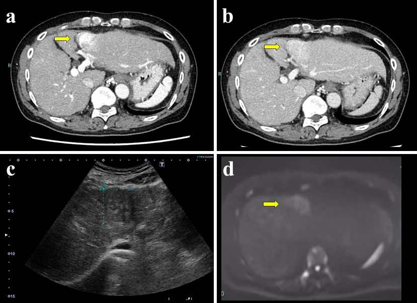
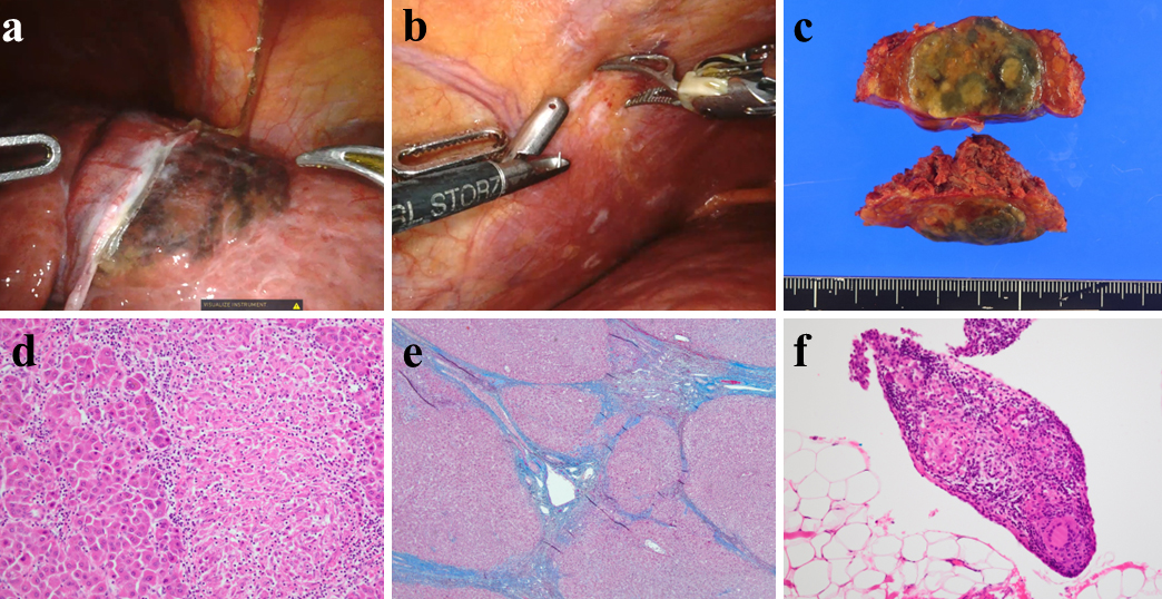
A 62-year-old patient with lung sarcoidosis presented with a 1.6-cm liver tumor. The patient reported no history of alcohol consumption or viral hepatitis. During the 3-year follow-up, the liver biopsies revealed hepatic sarcoidosis with no malignancy. However, the tumor eventually grew to 4.0 cm; radiographic findings are presented in Figure 1. The tumor was diagnosed as hepatocellular carcinoma (HCC). Consequently, robotic hepatectomy was performed, and intraoperative and pathological findings are demonstrated in Figure 2. Finally, the tumor was diagnosed as HCC associated with hepatic sarcoidosis.
Reportedly, the incidence of hepatic sarcoidosis is 4.2% in patients with sarcoidosis (1). Nonetheless, few studies have reported sarcoidosis associated with HCC (2), (3). Herein, the liver cirrhosis occurred because of hepatic sarcoidosis, further developing HCC. In summary, this report describes the clinical images of an extremely rare case of HCC associated with hepatic sarcoidosis.
None
YN and KT wrote the draft. All authors contributed equally to the manuscript. All authors edited, read, and approved the final manuscript.
Not applicable
Written informed consent was obtained from the patient for the publication of this report and any accompanying images.
Graf C, Arncken J, Lange CM, et al. Hepatic sarcoidosis: clinical characteristics and outcome. JHEP Rep. 2021;3(6):100360.
Ogata S, Horio T, Sugiura Y, et al. Sarcoidosis-associated hepatocellular carcinoma. Acta Med Okayama. 2010;64(6):407-10.
Arai T, Akita S, Sakon M, et al. Hepatocellular carcinoma associated with sarcoidosis. Int J Surg Case Rep. 2014;5(8):562-5.
