Corresponding author: Yusuke Namba, d5w8y.nmb@gmail.com
DOI: 10.31662/jmaj.2025-0033
Received: January 20, 2025
Accepted: February 13, 2025
Advance Publication: March 28, 2025
Published: April 28, 2025
Cite this article as:
Namba Y, Tokioka K, Kawai Y. Venous Thrombus Calcification over Time. JMA J. 2025;8(2):627-629.
Key words: thrombus, calcification, inferior vena cava, left common iliac vein, magnetic resonance imaging
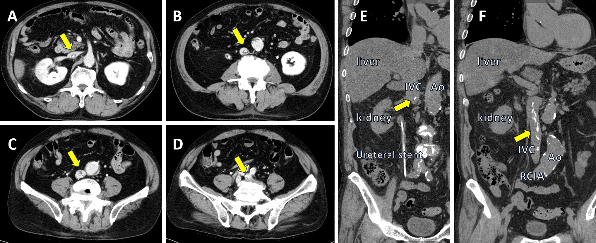
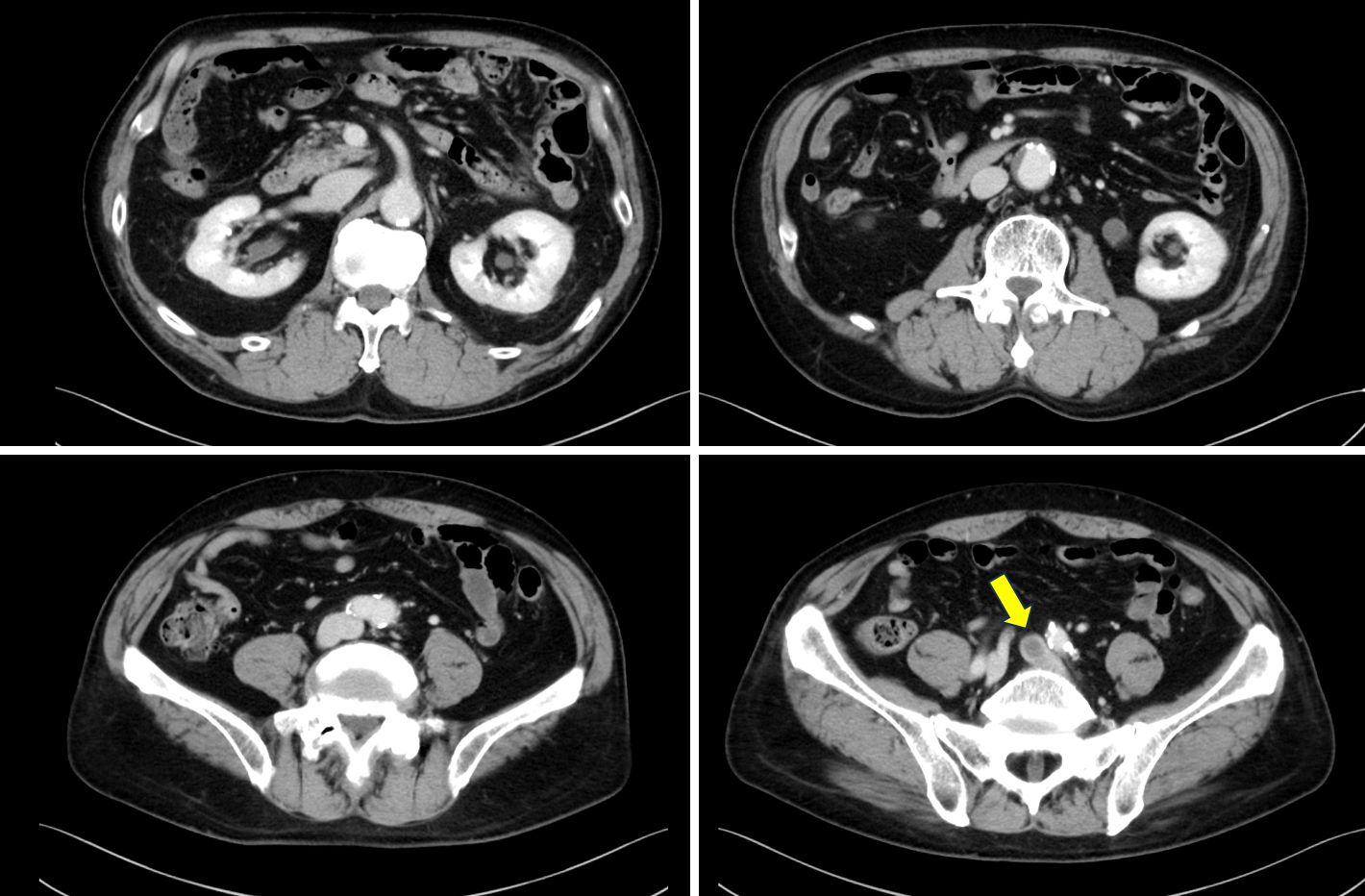
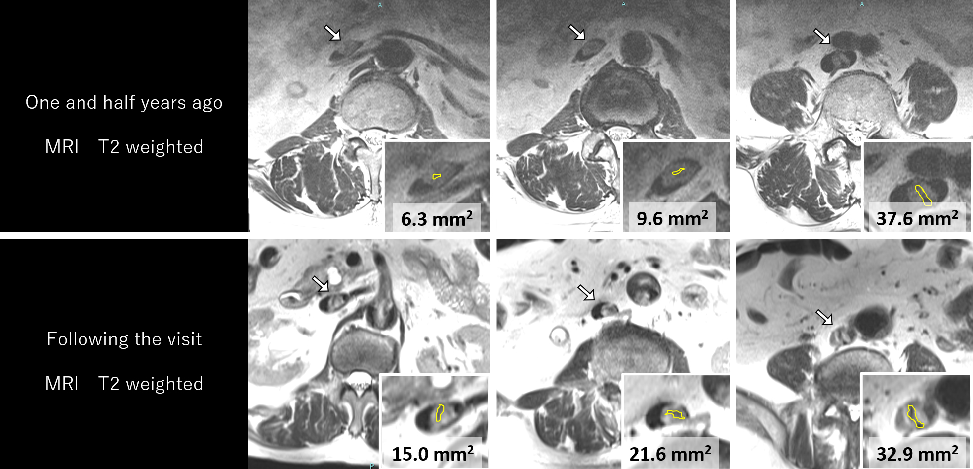
A man in his 60s with obstructive pyelonephritis due to upper urinary tract urolithiasis diagnosed 7 years earlier and no thrombophilia risk presented with left lower-extremity edema. Contrast-enhanced computed tomography (CeCT) revealed a calcified thrombus extending from the inferior vena cava to the left common iliac vein (LCIV; Figure 1). A CeCT performed 4.5 years prior showed a non-calcified thrombus in the LCIV (Figure 2), and no anticoagulants were prescribed. Magnetic resonance imaging (MRI) performed 18 months prior showed a low-intensity area in an intermediate-intensity area. MRI performed on the day following the visit demonstrated that the low-intensity area had increased over time (Figure 3). His edema resolved following rivaroxaban administration, which was continued thereafter.
A previous report demonstrated inflammatory infiltrate and fibrotic evolution in pulmonary artery thrombi over time (1).
However, no study has assessed non-calcified thrombi that calcified over time. We believe this case is significant because it shows that an organized thrombus can calcify. Clinicians should treat deep vein thrombosis formation with anticoagulants to prevent thrombi formation and calcification.
None
Yusuke Namba and Koji Tokioka managed the patient. Yusuke Namba wrote the original manuscript. Koji Tokioka and Yusuke Kawai reviewed and supervised the manuscript.
IRB approval was not required for this report.
Consent to publish the details of the present case was obtained from the patient.
Mansueto G, Costa D, Capasso E, et al. The dating of thrombus organization in cases of pulmonary embolism: an autopsy study. BMC Cardiovasc Disord. 2019;19(1):250.
