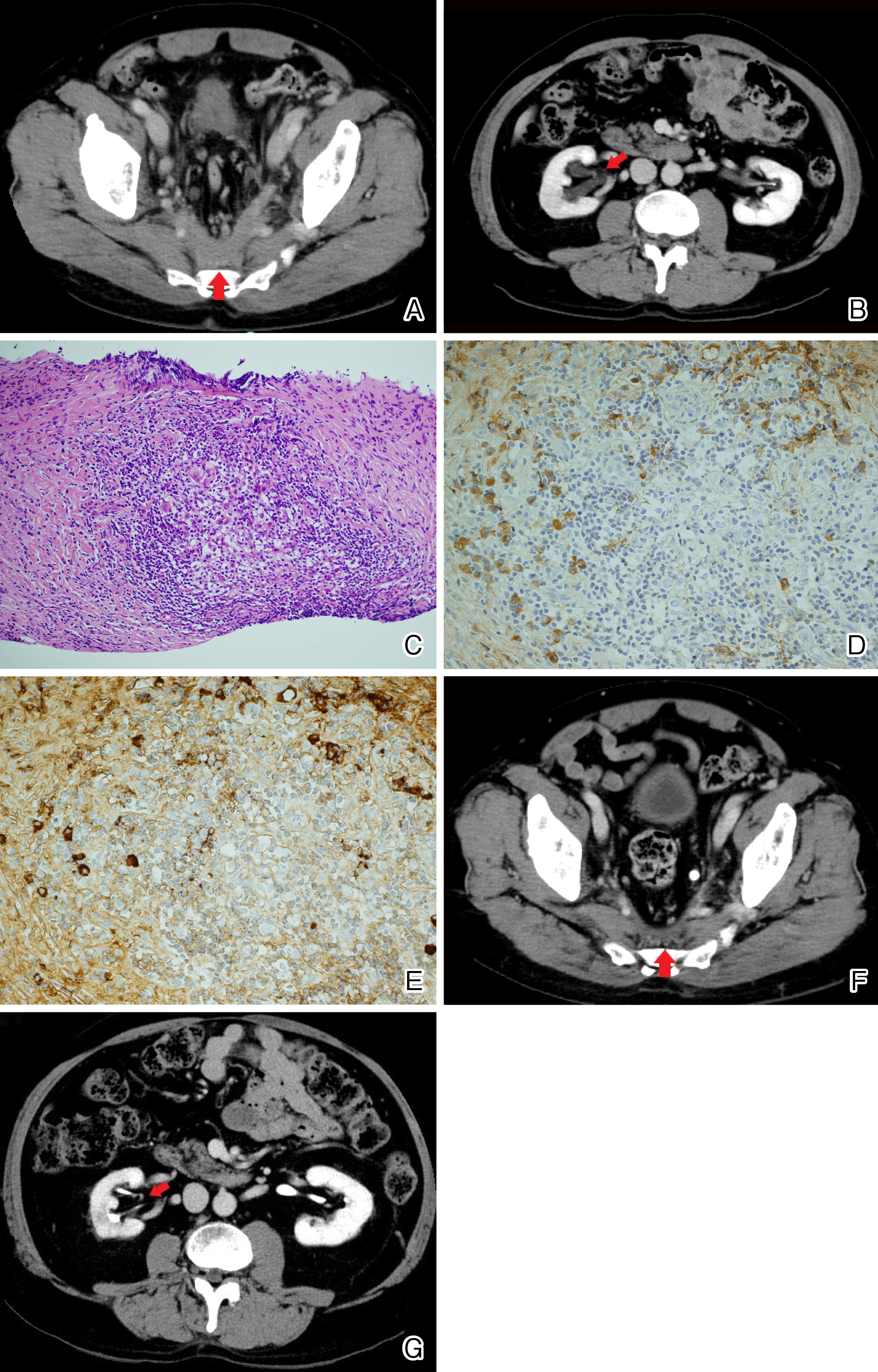
Figure 1. (A, B) Contrast-enhanced computed tomography scans before treatment showing a mass (red arrow) occupying the caudal retroperitoneal area surrounding the pelvic arteries and veins (A) and an enlarging mass that suppressed the right ureter, causing right-side hydronephrosis (red arrow) (B).
(C, D, E) Hematoxylin and eosin staining of core needle biopsy specimens taken from retroperitoneal mass showing stromal fibrosis with dense infiltration of normal lymphocytes and plasma cells. (magnification, × 200) (C). Immunohistochemical staining showing the infiltration of IgG-positive plasma cells (magnification, × 400) (D). The number of IgG4-positive plasma cells per high-power field was 30 (magnification, × 400) (E). The ratio of IgG4-positive plasma cells/IgG-positive cells was approximately 60%.
(F, G) After 2 months of treatment with oral prednisolone and azathioprine, the mass shrank (red arrow) (F) and the hydronephrosis subsequently improved (red arrow) (G).
From: Unilateral Leg Edema and Hydronephrosis in IgG4-Related Retroperitoneal Fibrosis