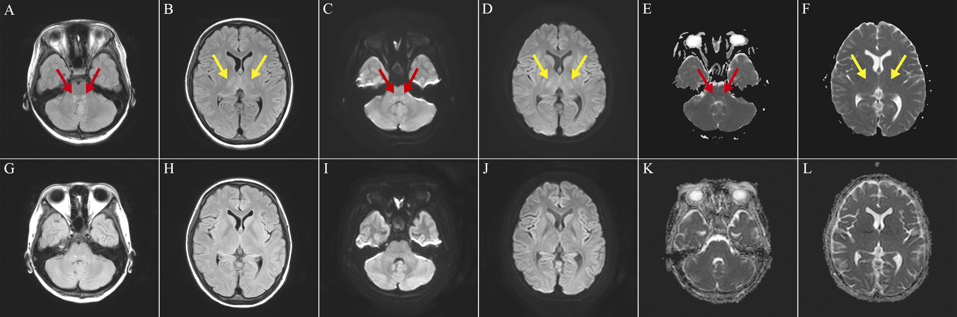
Figure 1. (A-F) Axial brain magnetic resonance imaging (MRI) of the patient showed bilateral symmetrical hyperintensities in the dorsal pontine base (red arrows) and thalamus (yellow arrows) on day 5 of hospitalization (A and B, T2-fluid-attenuated inversion recovery [FLAIR]; C and D, diffusion-weighted imaging; and E and F, apparent diffusion coefficient maps). (G-L) MRI signal changes in the pons and thalamus improved on day 12 of hospitalization (G and H, T2-FLAIR; I and J, diffusion-weighted imaging; and K and L, apparent diffusion coefficient maps).
From: Brain Magnetic Resonance Imaging Findings of Shiga Toxin-producing Escherichia coli Hemolytic Uremic Syndrome-associated Encephalopathy