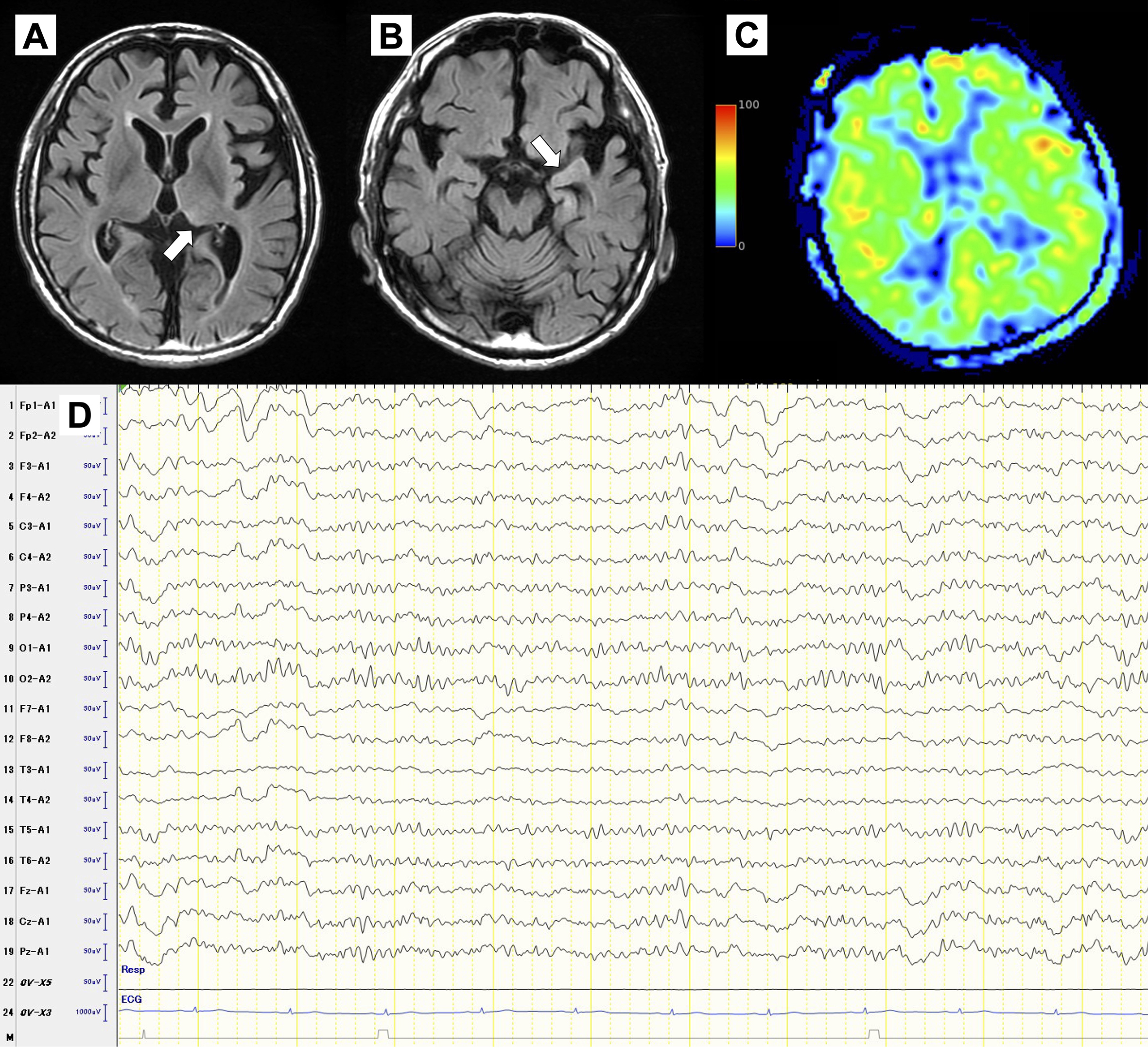
Figure 1. (A) and (B) Diffusion-weighted images (DWI) on initial presentation demonstrating increased signal intensity in the left pulvinar (A) and temporal tip (B). (C) Arterial spin labeling (ASL) on initial presentation demonstrated increased signal intensity in the left hemisphere. (D) Electroencephalography (EEG) on day 3, showed lateralized periodic discharges in the left frontal region.
From: Subacute Encephalopathy and Seizures in Alcoholics Syndrome
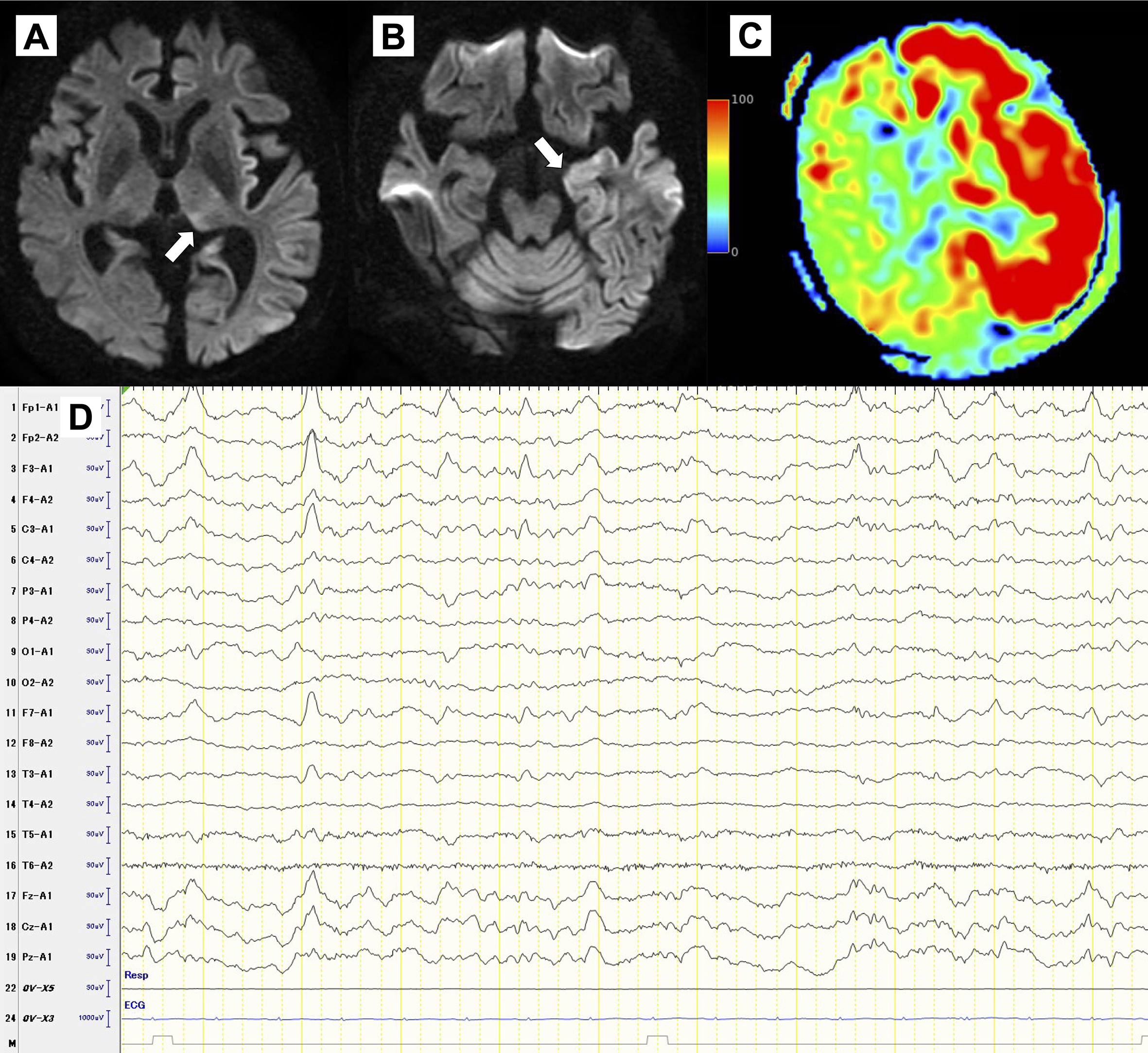
Figure 2. (A) and (B) Fluid-attenuated inversion recovery (FLAIR) on day 8, showing resolving hyperintensity in the left pulvinar (A) and temporal tip (B). (C) Arterial spin labeling (ASL) on day 8, demonstrated no increased signal intensity. (D) Electroencephalography (EEG) on Day 50, showed resolved lateralized periodic discharges.
From: Subacute Encephalopathy and Seizures in Alcoholics Syndrome