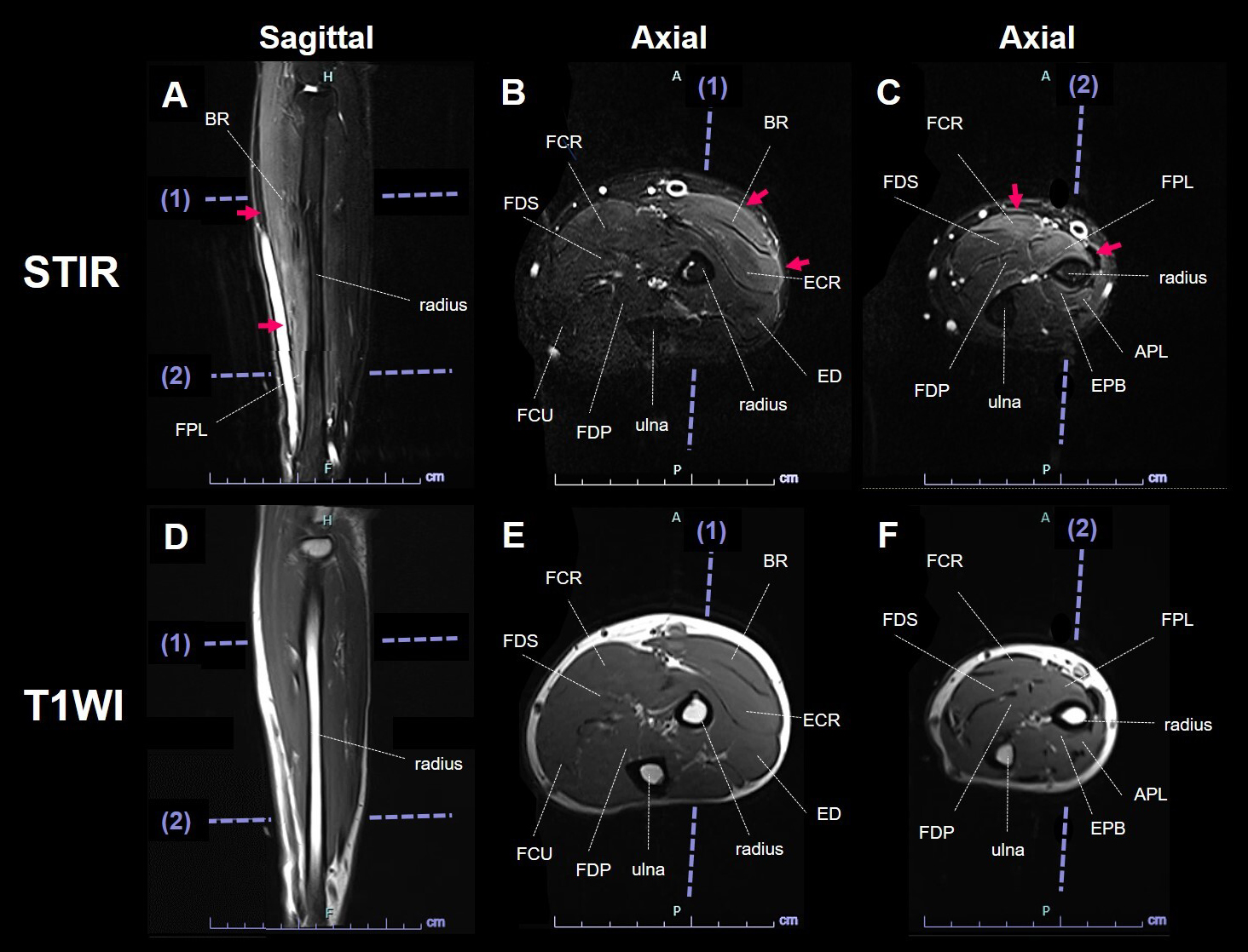
Figure 2. MRI of the left forearm.
STIR sagittal image (A) and axial images (B, C) show hyperintensity in the muscle and fascia in the BR, ECR, FCR, and FPL muscles (A-C, arrows). The FDP muscle appeared intact. T1-weighted images show normal findings (D-F).
APL: abductor pollicis longus; BR: brachioradialis; ECR: extensor carpi radialis longus and brevis; ED: extensor digitorum; EPB: extensor pollicis brevis; FCR: flexor carpi radialis; FCU: flexor carpi ulnaris; FDP: flexor digitorum profundus; FDS: flexor digitorum superficialis; FPL: flexor pollicis longus; MRI: magnetic resonance imaging; STIR: short-tau inversion recovery; TIWI: T1-weighted image.
From: Forearm Magnetic Resonance Imaging Findings in Epidemic Myalgia