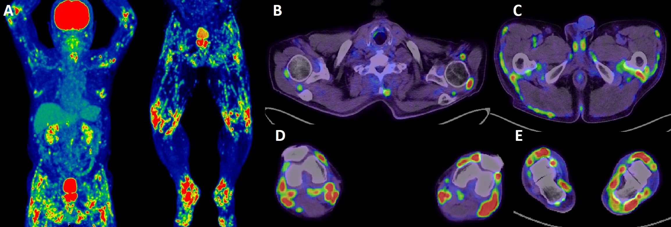
Figure 1. Imaging findings of the patient.
This panel presents 18F-fluorodeoxyglucose positron emission tomography/computed tomography showing accumulation of fluorodeoxyglucose in numerous muscle-tendon attachment sites. (A) whole body, (B) shoulder region, (C) lumbar region, (D) knee region, and (E) foot region. SUVmean ± SD of each site: attachment sites of subscapular fossa and subscapularis muscle, 3.25 ± 0.86; attachment sites of the clavicle and subclavian muscle, 2.00 ± 0.56; attachment sites of the humeral head and supraspinatus muscle, 6.85 ± 2.38; attachment sites of the humeral head and biceps brachii tendon, 4.68 ± 1.45; attachment sites of femur and gluteus minimus, 2.54 ± 0.31; attachment sites of femur and gluteus medius, 2.80 ± 0.45; attachment sites of coccyx and gluteus maximus, 5.01 ± 0.84; attachment sites of the coccyx and iliac muscle, 4.36 ± 0.71; attachment sites of the coccyx and obturatorius externus muscle, 3.46 ± 0.73; and tendon-to-bone attachment sites of knees, 12.70 ± 2.44.
SD: standard deviation; SUVmean: mean standardized uptake value.
From: Diffuse Polyenthesitis after Intravesical Bacillus Calmette-Guerin Therapy