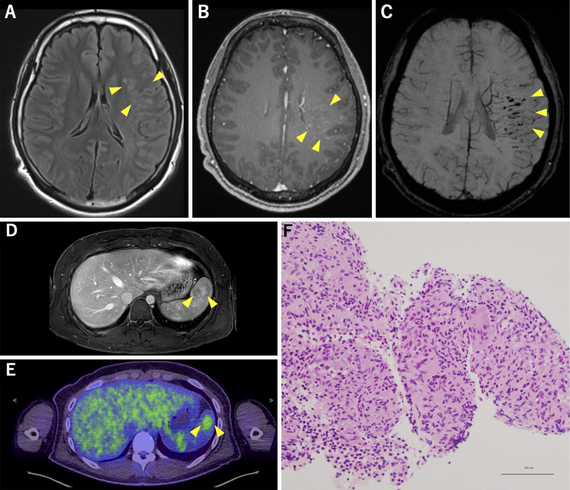
Figure 1. Brain MRI reveals hyperintense lesions in the left inferior frontal gyrus, superior temporal gyrus, and supramarginal gyrus on axial FLAIR (A, yellow arrowheads), and gadolinium-enhanced T1-weighted imaging shows linear contrast enhancement along medullary veins (B, yellow arrowheads). Susceptibility-weighted imaging reveals dilated medullary veins and multiple hypointense areas indicative of microbleeds (C, yellow arrowheads). Abdominal MRI reveals mild splenomegaly and a focal lesion with heterogeneous contrast enhancement during portal venous phase (D, yellow arrowheads), and 18F-FDG-PET/CT shows abnormal uptake corresponding to this splenic region, with a an SUV max of 3.1 (E, yellow arrowheads). A splenic biopsy confirms clusters of epithelioid cells without caseous necrosis (F).
18F-FDG-PET/CT: 18F-fluorodeoxyglucose positron emission tomography/computed tomography; FLAIR: fluid-attenuated inversion recovery; MRI: magnetic resonance imaging; SUV max: maximal standardized uptake value.
From: Neurosarcoidosis Limited to the Central Nervous System and Spleen, Presenting with Episodic Nonfluent Aphasia