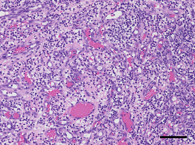
Figure 3. Microscopic findings of the specimen. The lesion was lined by stratified squamous epithelium, and consisted of prominent hypervascularization and fibrous connective tissues with inflammatory cell infiltration. Hematoxylin and eosin staining, bar 100 μm, ×200.
From: Pregnancy Epulis