Corresponding author: Yoshitsugu Chigusa, chigusa@kuhp.kyoto-u.ac.jp
DOI: 10.31662/jmaj.2022-0156
Received: August 17, 2022
Accepted: November 24, 2022
Advance Publication: February 27, 2023
Published: April 14, 2023
Cite this article as:
Chigusa Y, Okuda R, Teratani Y, Matsuzaka S, Mandai M. Pregnancy Epulis. JMA J. 2023;6(2):206-208.
Key words: epulis, granuloma, pregnancy
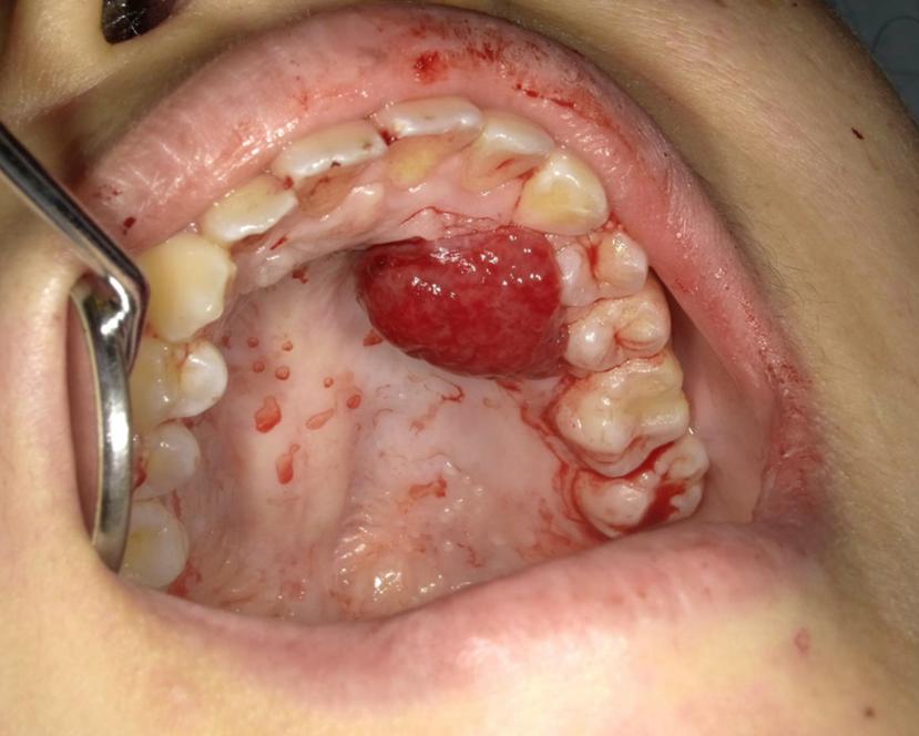
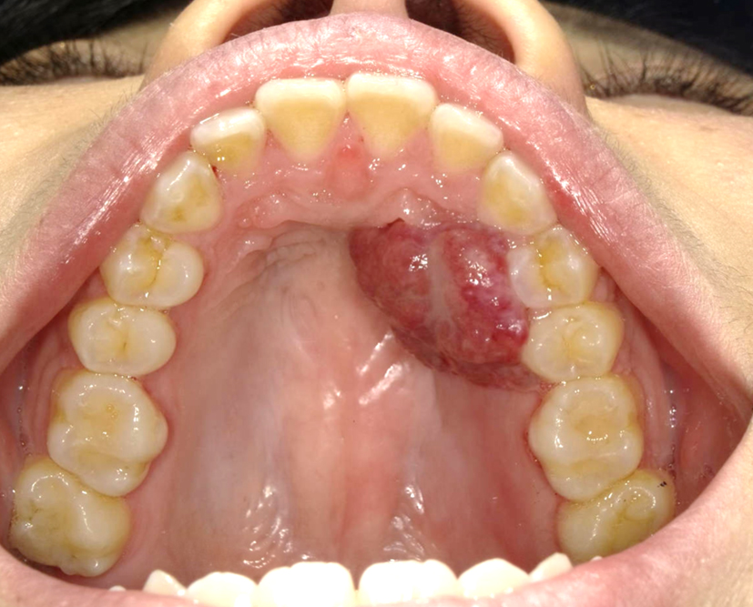
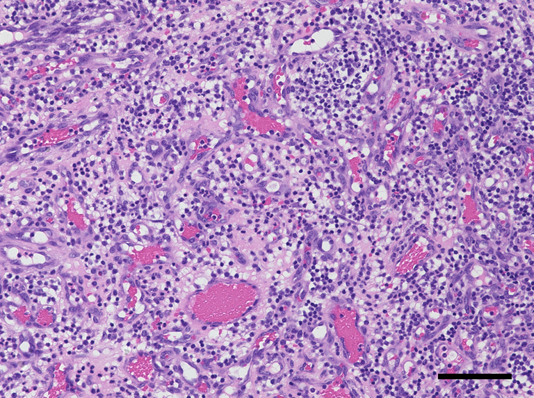
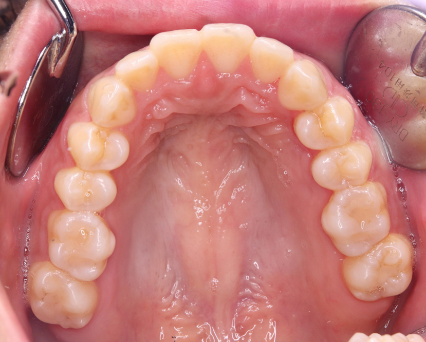
A 29-year-old primigravida was presented with intraoral bleeding at the 38+0/7 weeks of gestation. A granuloma-like mass of about 23 mm with bleeding was found in left maxillary gingiva (Figure 1). This lesion had arisen during her pregnancy and had grown rapidly and repeatedly bled in the past month. The bleeding stopped after 20 min of pressure by the dentist. The same day, she underwent emergent cesarean section due to nonreassuring fetal status. Thirty-six days postpartum, the tumor was resected (Figure 2). Microscopically, the lesion was lined by stratified squamous epithelium and comprised prominent hypervascularization and fibrous connective tissues with inflammatory cell infiltration, indicating pregnancy epulis (Figure 3). The postoperative course was uneventful (Figure 4). Pregnancy epulis is a hyperplastic lesion originating from oral mucosa (1), (2). The excision is not recommended during pregnancy because of the risk of heavy bleeding and the possibility of regression after delivery. However surgical resection should be considered if it does not shrink even after childbirth. Complete excision and subsequent maintenance of oral hygiene are important to prevent recurrence (3).
None
We would like to thank Editage (www.editage.com) for English language editing.
RO, YT, and SM contributed to patient care, and collected patient’s data and images. YC wrote the manuscript, and the other authors revised it. MM supervised the entire process.
This study did not require IRB approval.
The written informed consent was obtained from the patient to publish this case, including pictures.
Anneroth G, Sigurdson A. Hyperplastic lesions of the gingiva and alveolar mucosa. A study of 175 cases. Acta Odontol Scand. 1983;41(2):75-86.
Cardoso JA, Spanemberg JC, Cherubini K, et al. Oral granuloma gravidarum: a retrospective study of 41 cases in Southern Brazil. J Appl Oral Sci. 2013;21(3):215-8.
StatPearls [Internet]. Sarwal P, Lapumnuaypol K. StatPearls. Treasure Island (FL); 2022, Pyogenic Granuloma. [cited 2022 Oct 26]. Available from: https://www.ncbi.nlm.nih.gov/books/NBK556077/.
