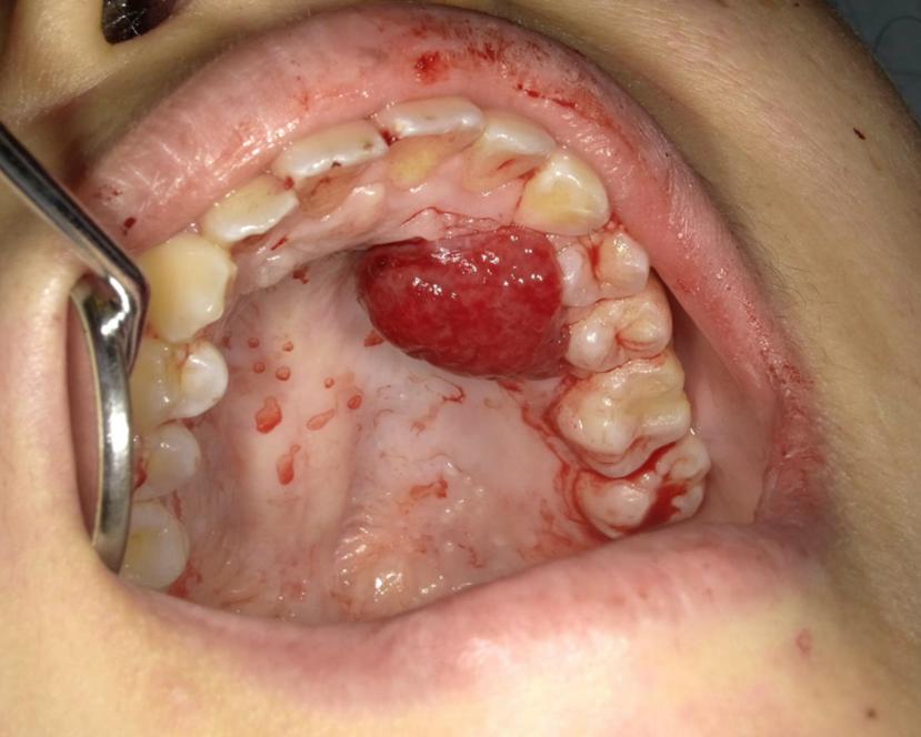
Figure 1. Intraoral image at the time of admission. A granuloma-like mass with irregular surface and bleeding was found.

From: Pregnancy Epulis

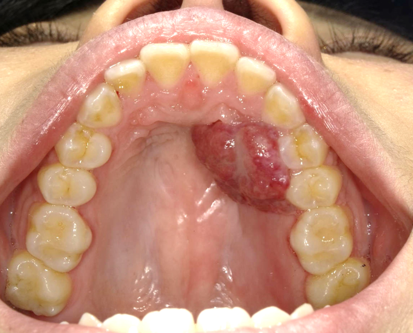
Figure 2. Intraoral image at the time of surgery. The lesion did not shrink spontaneously after childbirth.

From: Pregnancy Epulis

Figure 3. Microscopic findings of the specimen. The lesion was lined by stratified squamous epithelium, and consisted of prominent hypervascularization and fibrous connective tissues with inflammatory cell infiltration. Hematoxylin and eosin staining, bar 100 μm, ×200.

From: Pregnancy Epulis
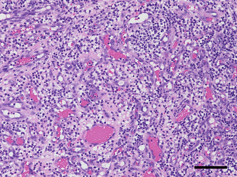
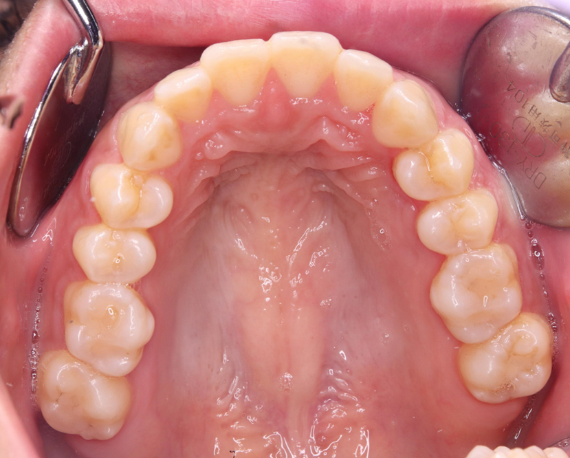
Figure 4. Intraoral image at one month postoperatively. Wound healing was satisfactory.

From: Pregnancy Epulis

PAGE TOP