Corresponding author: Masahiro Ito, soxboston@yahoo.co.jp
DOI: 10.31662/jmaj.2022-0199
Received: October 31, 2022
Accepted: November 25, 2022
Advance Publication: March 6, 2023
Published: April 14, 2023
Cite this article as:
Ito M, Sato T, Nagai T. Epiploic Appendagitis: A Rare Cause of Acute Abdominal Pain. JMA J. 2023;6(2):209-210.
Key words: epiploic appendagitis, computed tomography, acute abdomen
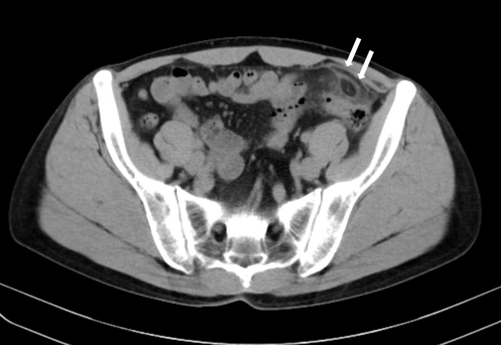
A previously healthy 48-year-old man presented with sudden-onset lower left abdominal pain, which persisted for 3 days. On physical examination, he had left lower quadrant tenderness. Laboratory results showed an increased white blood cell count (9.8 × 1000/μl) and increased C-reactive protein (1.8 mg/dL). Abdominal computed tomography (CT) revealed a fat-density ovoid structure with a high-density rim adjacent to the sigmoid colon and surrounding inflammatory fat, which was consistent with epiploic appendagitis (Figure 1). The patient was treated with a nonsteroidal anti-inflammatory drug and his pain resolved after 1 week. Epiploic appendagitis has nonspecific clinical findings (1), and abdominal CT is of critical importance to properly diagnose this disease and distinguish it from other surgical emergencies responsible for acute abdomen (2). This condition is basically a self-limiting disease; however, laparoscopic appendage excision may be required for recurrent cases (1). Epiploic appendagitis is a rare condition but should be considered in patients with acute abdominal pain.
None
MI wrote the first draft and managed the submission processes. All authors (1) made substantial contributions to the study concept and data analysis/interpretation; (2) drafted the manuscript and revised it critically for important intellectual content; (3) approved the final version of the manuscript to be published; and (4) agreed to be accountable for all aspects of the work.
We have obtained informed consent for the publication of this case report.
Giannis D, Matenoglou E, Sidiropoulou MS, et al. Epiploic appendagitis: pathogenesis, clinical findings and imaging clues of a misdiagnosed mimicker. Ann Transl Med. 2019;7(24):814-22.
Giambelluca D, Cannella R, Caruana G, et al. CT imaging findings of epiploic appendagitis: an unusual cause of abdominal pain. Insights Imaging. 2019;10(1):26-35.
