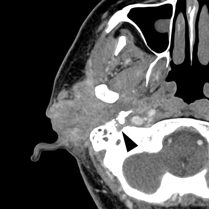
Figure 1. Contrast-enhanced computed tomography shows a contrast-enhanced tumor extending from the right external auditory canal to the piriformis and invading the right facial nerve. The arrowhead indicates the tumor extending into the stylomastoid foramen.
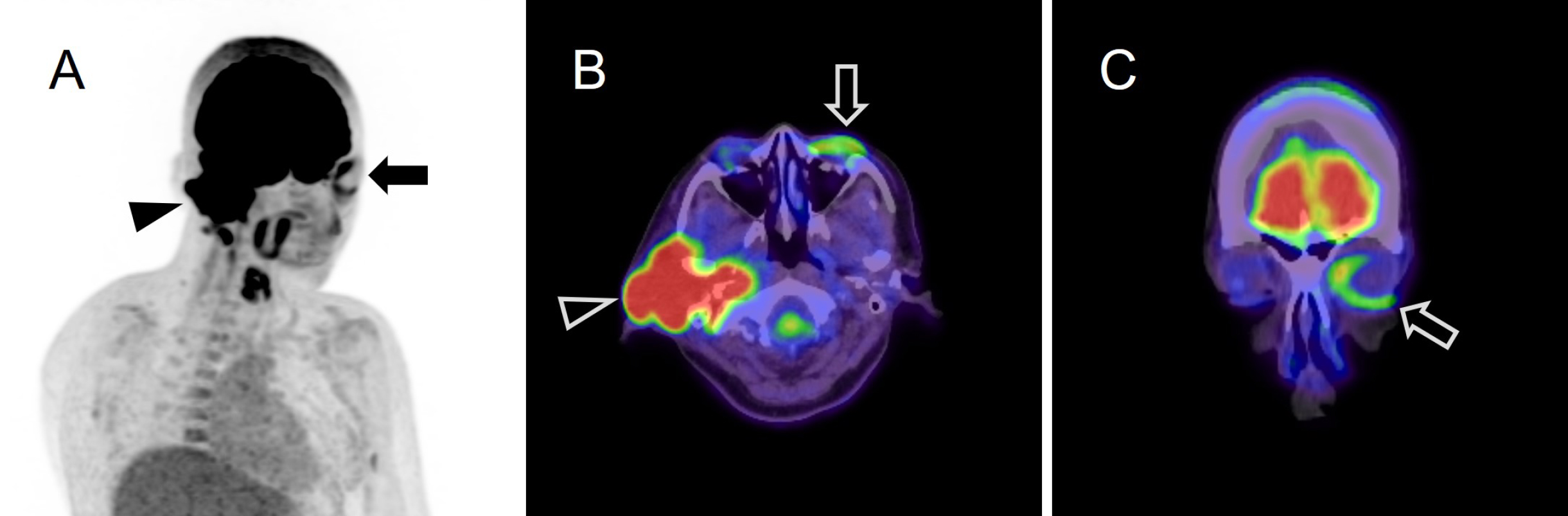
Figure 2. Positron emission tomography/computed tomography showing intense 18F-FDG uptake associated with right external auditory canal cancer (A, B: arrow heads) and increased 18F-FDG uptake (maximum standardized uptake value: 3.08) in the left orbicularis oculi muscle on the unaffected side (A, B, C: arrows). 18F-FDG: 18F-fluorodeoxyglucose.