Figure 1. Contrast-enhanced computed tomography (CT) images upon arrival and intraoperative findings.
(A) Coronal view showing an extensive thrombus in the main trunk of the portal vein (arrowhead).
(B) A poor contrast area within a section of the small intestine accompanied by intestinal wall edema and thickening (circles).
(C) Intraoperative findings showing bloody ascites, extensive necrosis of approximately 150 cm of the jejunum, and mesenteric edema.
From: Hybrid Surgery for Superior Mesenteric Vein Thrombosis: A Case Report
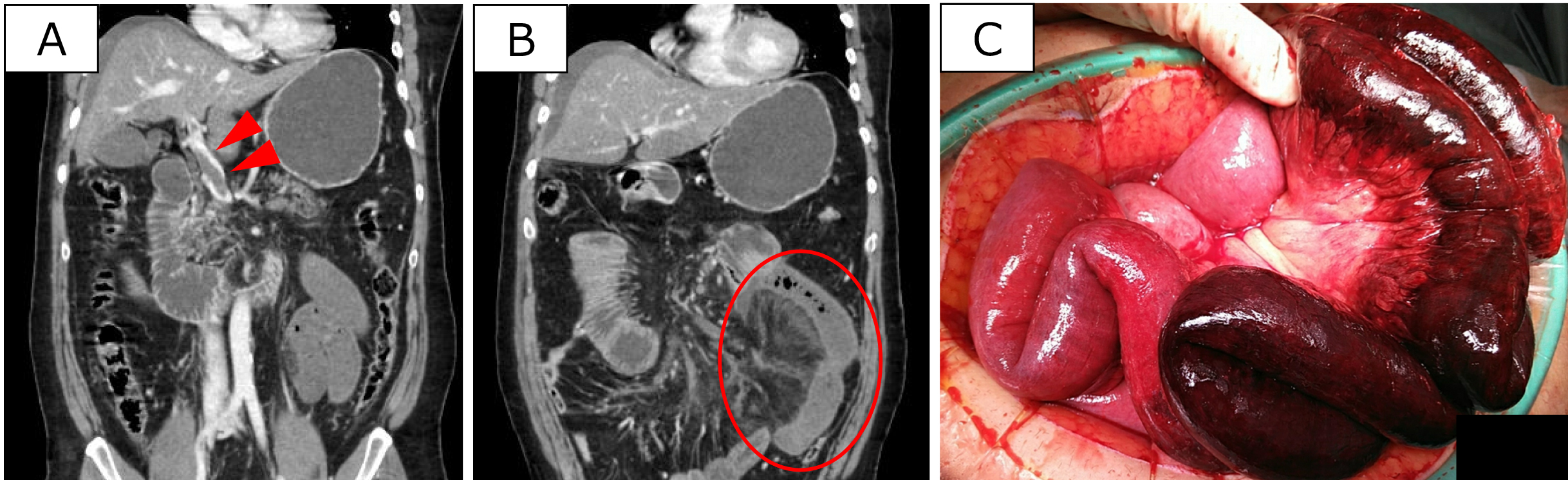
Figure 2. Endovascular thrombectomy of the portal vein.
(A1) Superior mesenteric venography shows a diffuse contrast defect. (A2) Thrombus removal from the portal vein using a stent retriever (arrowhead). (A3) Confirmatory portography showing improved blood flow, with residual thrombus remaining.
(B) The large thrombus volume is removed using the stent retriever.
From: Hybrid Surgery for Superior Mesenteric Vein Thrombosis: A Case Report
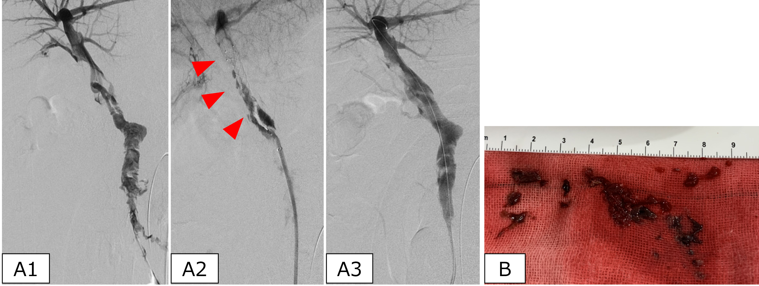
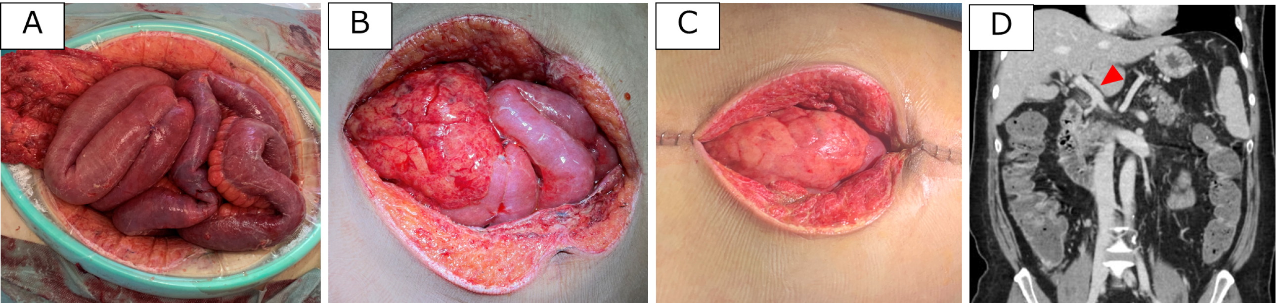
Figure 3. Bowel findings before and after thrombus retrieval.
Condition of the open abdominal wound: (A) day 4, (B) day 6, and (C) day 11.
Intestinal edema progressively improved, allowing for staged closure of the laparotomy wound.
(D) Follow-up contrast-enhanced CT on day 42, showing resolution of the intraportal thrombus (arrowhead).
From: Hybrid Surgery for Superior Mesenteric Vein Thrombosis: A Case Report