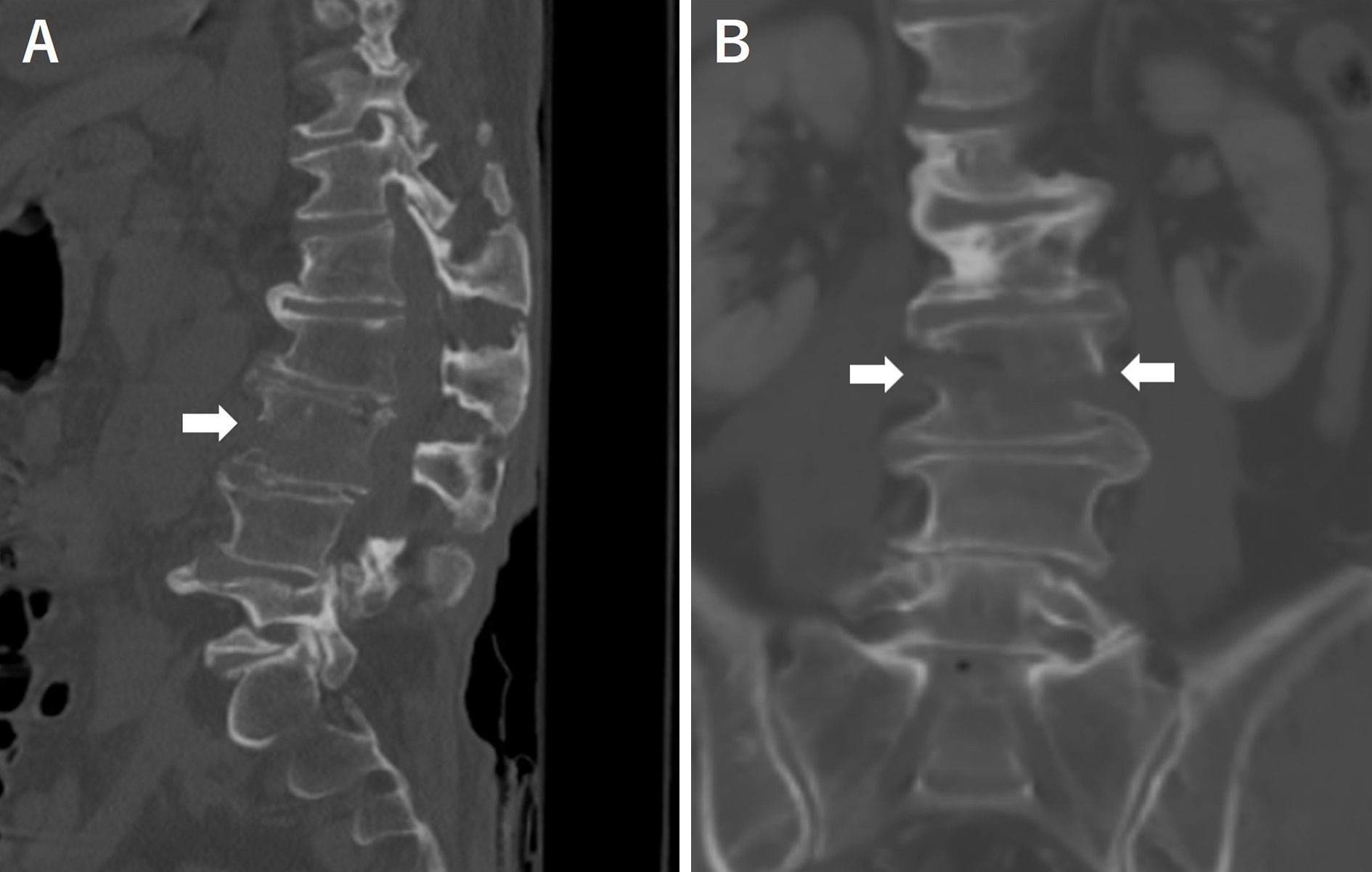
Figure 1. Sagittal (A) and coronal (B) slices of plain computed tomography revealed diffuse idiopathic skeletal hyperostosis and a fracture of the third lumbar vertebra.
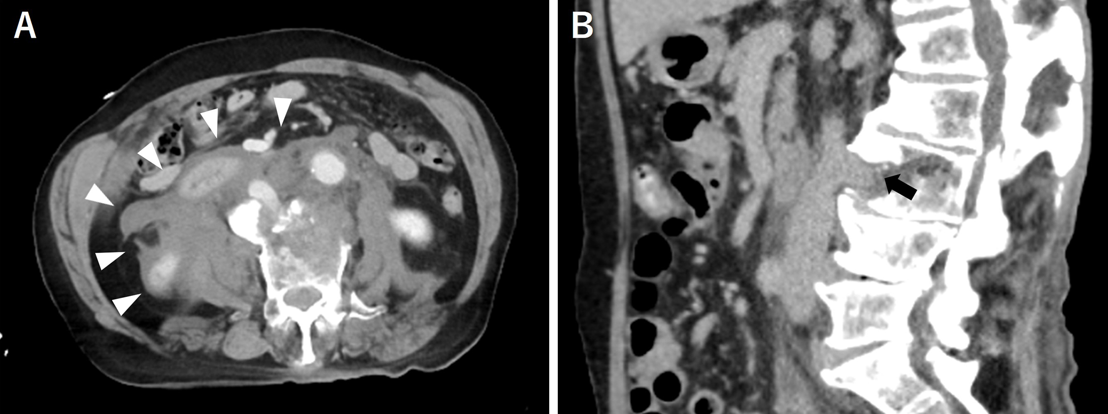
Figure 2. An axial slice (A) of the contrast-enhanced computed tomography revealed a large amount of hematoma in the abdominal cavity, whereas a sagittal slice (B) showed that the inferior vena cava was damaged and in contact with the fracture site.
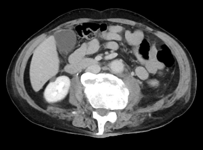
Figure 3. A contrast computed tomography scan taken 3 weeks after the injury showed that the hematoma almost disappeared, and there was no bleeding from the inferior vena cava.