Corresponding author: Yushi Sakamoto, bebenko19831210@gmail.com
DOI: 10.31662/jmaj.2024-0356
Received: November 11, 2024
Accepted: November 15, 2024
Advance Publication: January 31, 2025
Published: April 28, 2025
Cite this article as:
Sakamoto Y, Takemura H, Ozaki T, Sugiki D, Yamazaki D. Images of Inferior Vena Cava Injury Secondary to Lumbar Vertebral Fracture in Diffuse Idiopathic Skeletal Hyperostosis. JMA J. 2025;8(2):599-601.
Key words: inferior vena cava injury, diffuse idiopathic skeletal hyperostosis, vertebral fracture
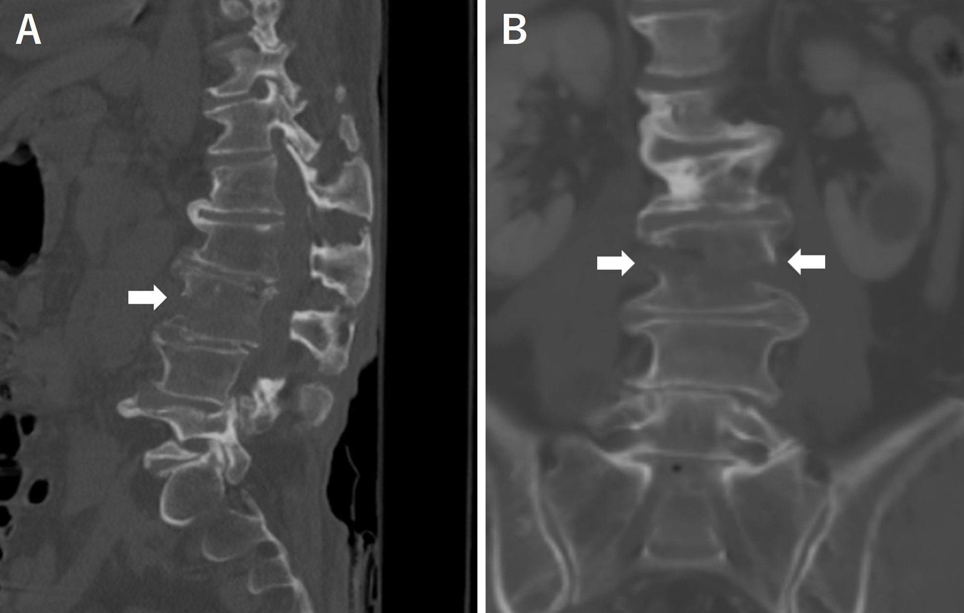
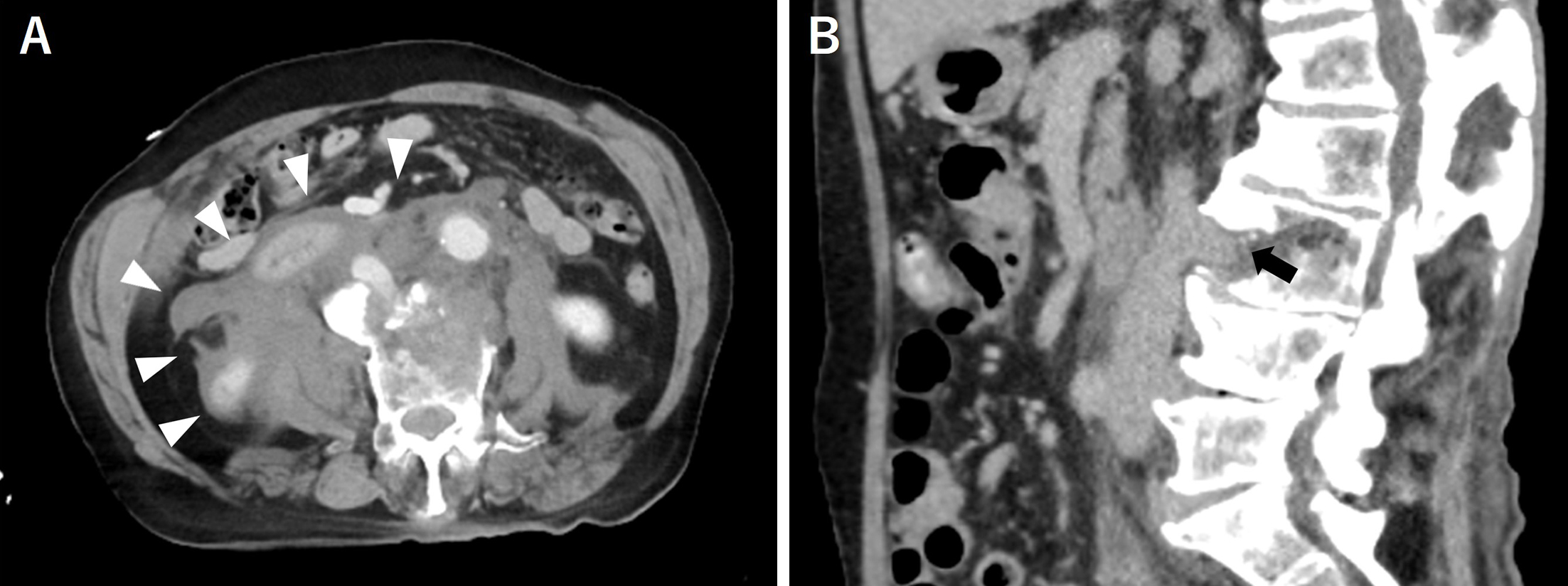
A 71-year-old man was hit by a car while crossing the road and was rushed to our hospital. A plain computed tomography (CT) scan revealed a vertebral fracture of the third lumbar vertebra associated with diffuse idiopathic skeletal hyperostosis (Figure 1A and 1B arrow). A subsequent contrast-enhanced CT scan revealed a large hematoma in the abdominal cavity (Figure 2A arrow head), and the inferior vena cava was trapped at the fracture site (Figure 2B arrow) (1).
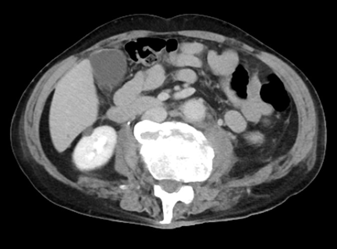
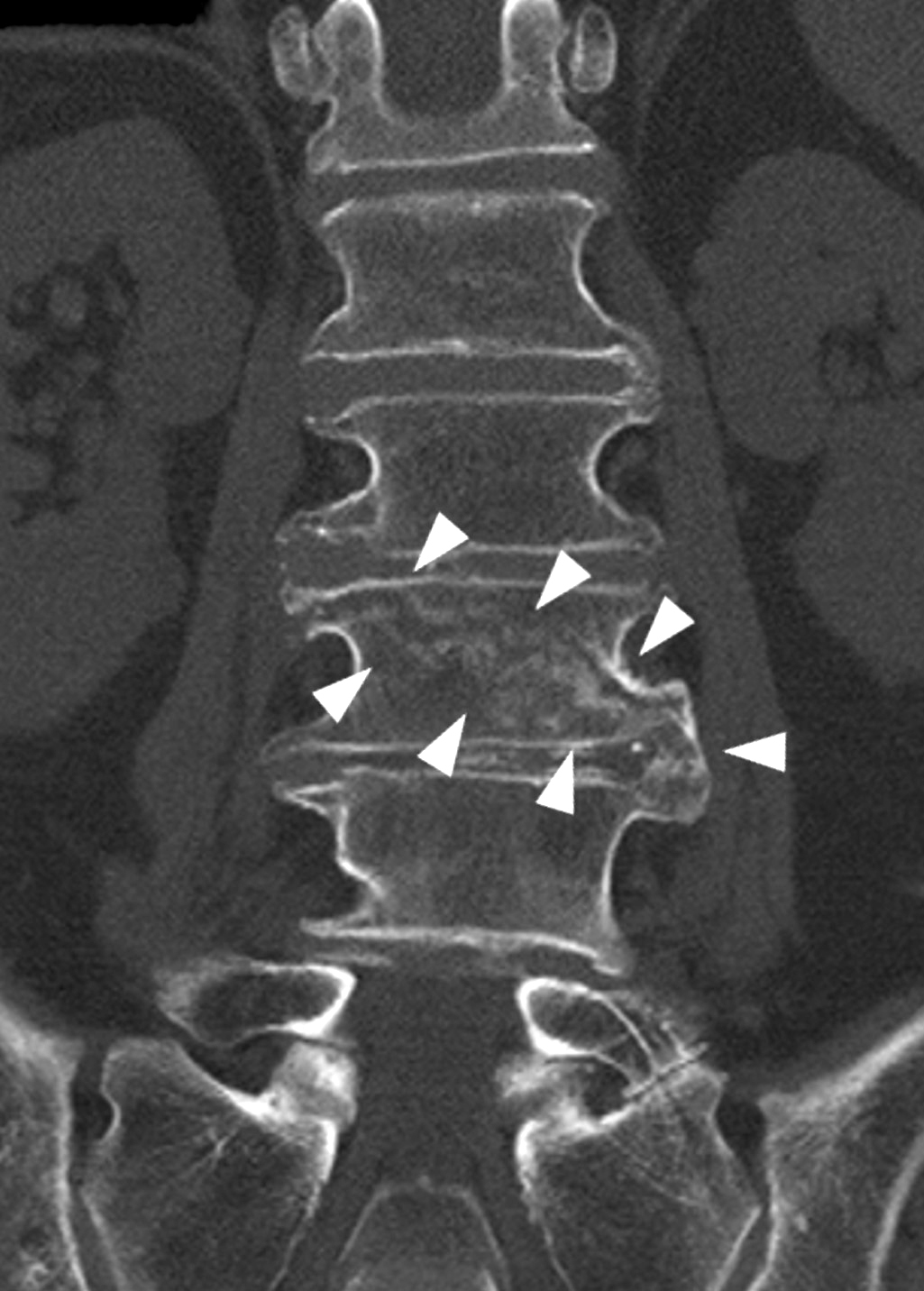
We considered surgery for the vertebral fracture but chose conservative treatment because of bleeding management issues. Three weeks after injury, the hematoma nearly disappeared, and bleeding in the inferior vena cava stopped (Figure 3). After 2 months of bed rest, a CT scan showed callus formation at the fracture site (Figure 4, arrowhead). Walking training was initiated with a corset. The patient started walking independently and was discharged 3 months after injury.
None
Yushi Sakamoto: care of patient and writing and editing manuscript
Hidetaka Takemura, Tomonori Ozaki, Dan Sugiki and Daisuke Yamazaki: care of patient
The patient understood and consented to the anonymous submission of the case report to a medical journal.
Eleti S, Roshen M, Griffiths M, et al. Imaging in traumatic injury to the inferior vena cava. Clin Radiol. 2021;76(10):787.e15-25.
