Figure 1. (a) Contrast-enhanced computed tomography images showing an early enhancement tumor at segment 3; the liver shape suggested a cirrhotic pattern. (b) Washout on delayed phase; (c) abdominal ultrasound showing mosaic architecture; (d) magnetic resonance imaging showing high signal intensity on diffusion-weighted images.
From: A Case of Hepatocellular Carcinoma Associated with Hepatic Sarcoidosis
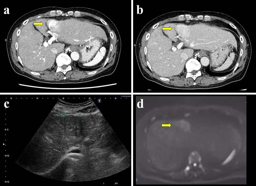
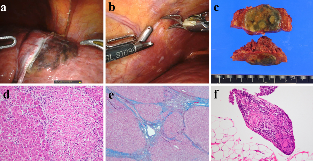
Figure 2. (a) Intraoperative findings showing a well-defined black color tumor on the segment 3 surface; (b) multiple white peritoneal nodules; (c) macroscopic findings showing a 3.2-cm green-yellowish tumor, capsulated confluent multinodular type; (d) microscopic findings showing moderately differentiated hepatocellular carcinoma and epithelioid granulomas; (e) background liver histology showing bridging fibrosis and regenerating nodules accompanied by the distortion of the liver lobules (f4); (f) epithelioid granulomas in a peritoneal nodule, suggesting peritoneal sarcoidosis.
From: A Case of Hepatocellular Carcinoma Associated with Hepatic Sarcoidosis