Figure 1. Preoperative T2-weighted MRI images (A, sagittal; B, axial) and CT scans (C, sagittal; D, axial) showing the dural sac severely compressed by a synovial cyst from the right facet at the L3/4 level (arrow, synovial cyst).

From: Full-endoscopic Removal of a Lumbar Synovial Cyst after Laminectomy for Lumbar Spinal Canal Stenosis
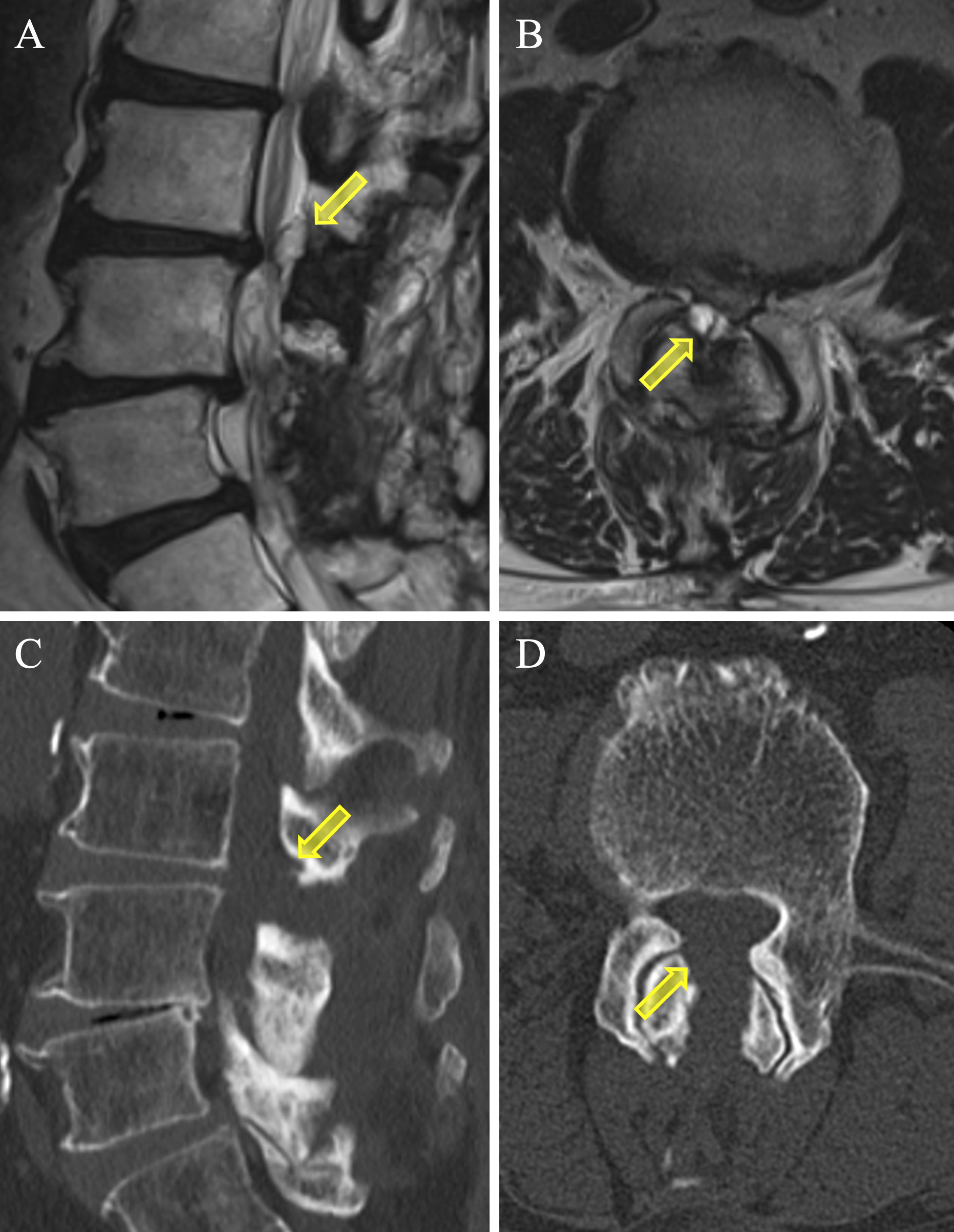
Figure 2. Intraoperative view with full-endoscopic spine surgery (FESS). The synovial cyst repeatedly shrank (A) and swelled (B) on the basis of respiratory fluctuations. The view (C) just before resection of the synovial cyst, and its removal, causing good pulsation of the dural sac (D) (arrowhead; synovial cyst).

From: Full-endoscopic Removal of a Lumbar Synovial Cyst after Laminectomy for Lumbar Spinal Canal Stenosis
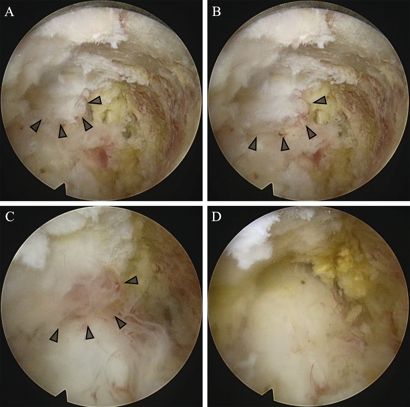
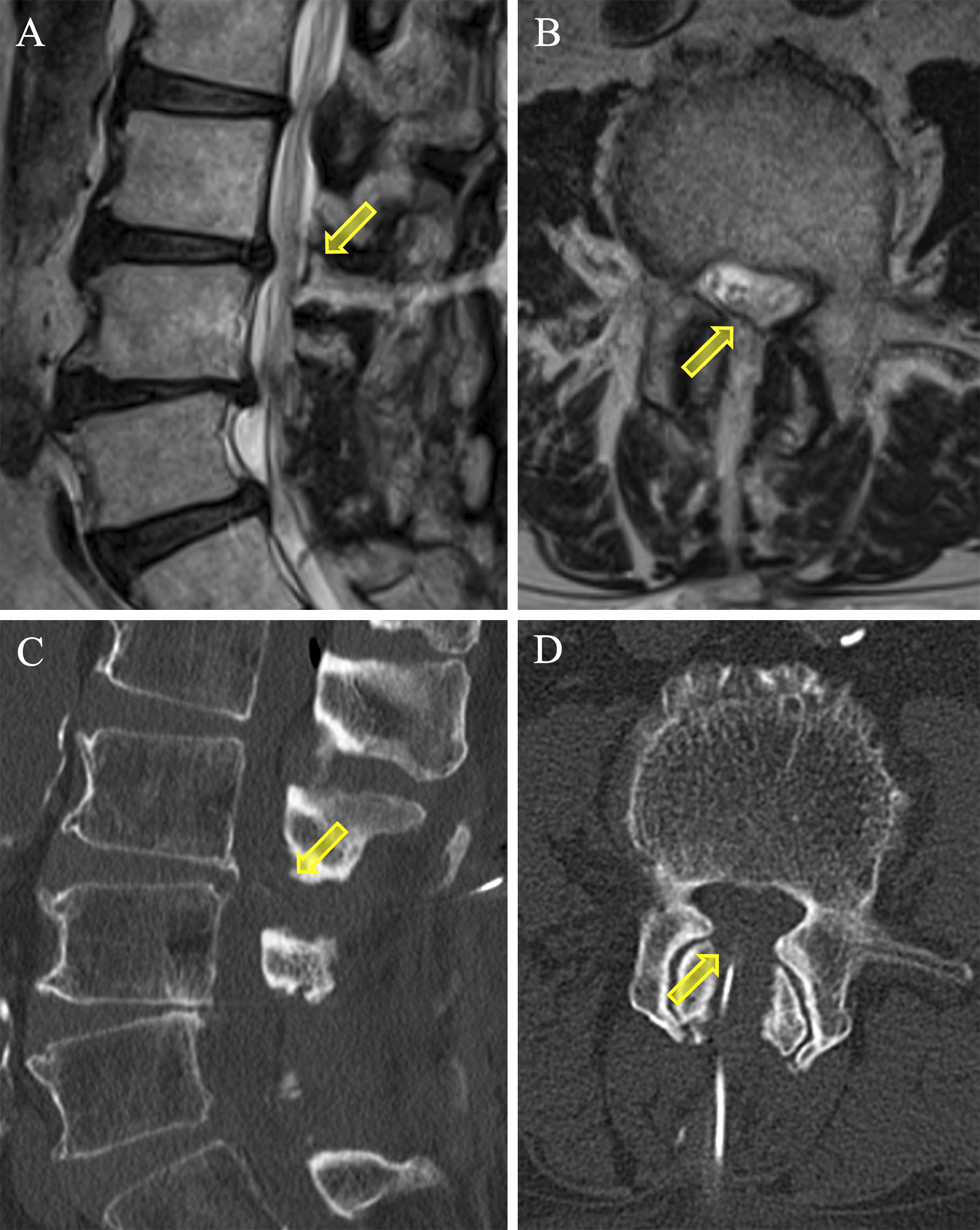
Figure 3. Postoperative T2-weighted MRI images (A; sagittal, B; axial) and CT scans (C; sagittal, D; axial) showing adequate decompression of the dural sac at the L3/4 level (arrow; disappearance of synovial cyst).
CT: computed tomography; MRI: magnetic resonance imaging.

From: Full-endoscopic Removal of a Lumbar Synovial Cyst after Laminectomy for Lumbar Spinal Canal Stenosis
PAGE TOP