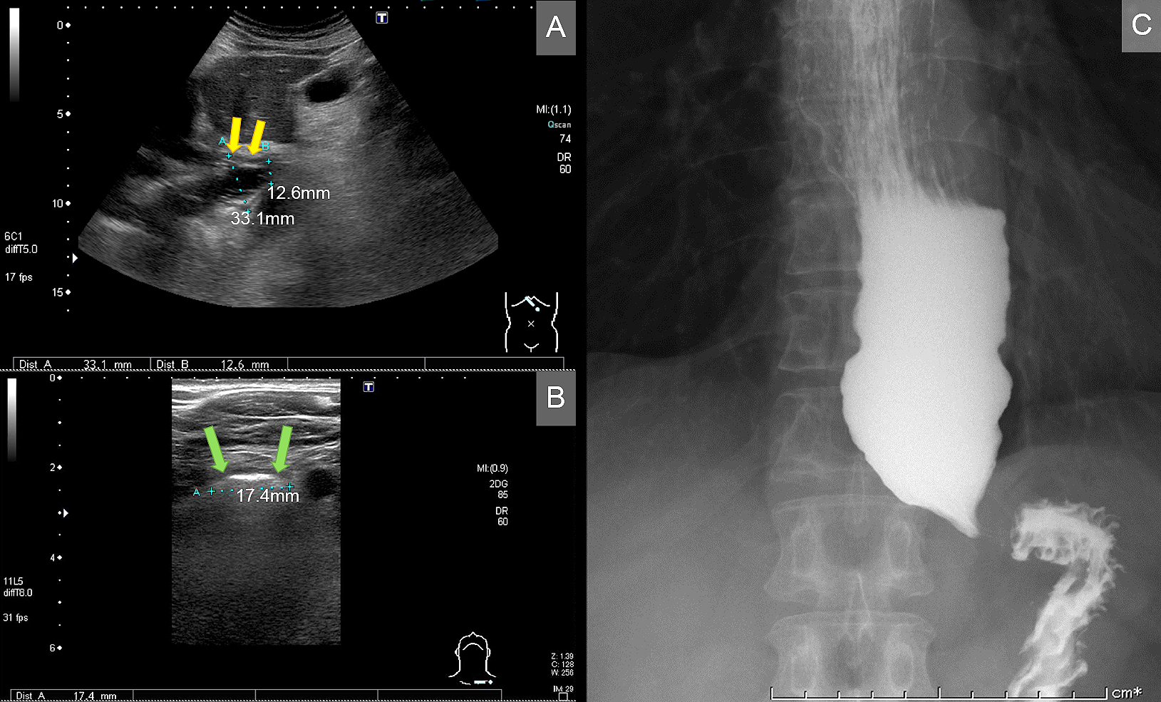
Figure. A transabdominal and neck ultrasonogram showing a dilated esophagus. (A) Significant dilatation and narrowing of the distal segment of the esophagus, with regular hypoechoic wall thickening (yellow arrow). (B) A neck ultrasonogram showing the dilated esophagus (green arrow). (C) A barium esophagogram showing the dilated esophagus, consistent with achalasia.
From: The Esophagus on Sight: Ultrasonography in Achalasia