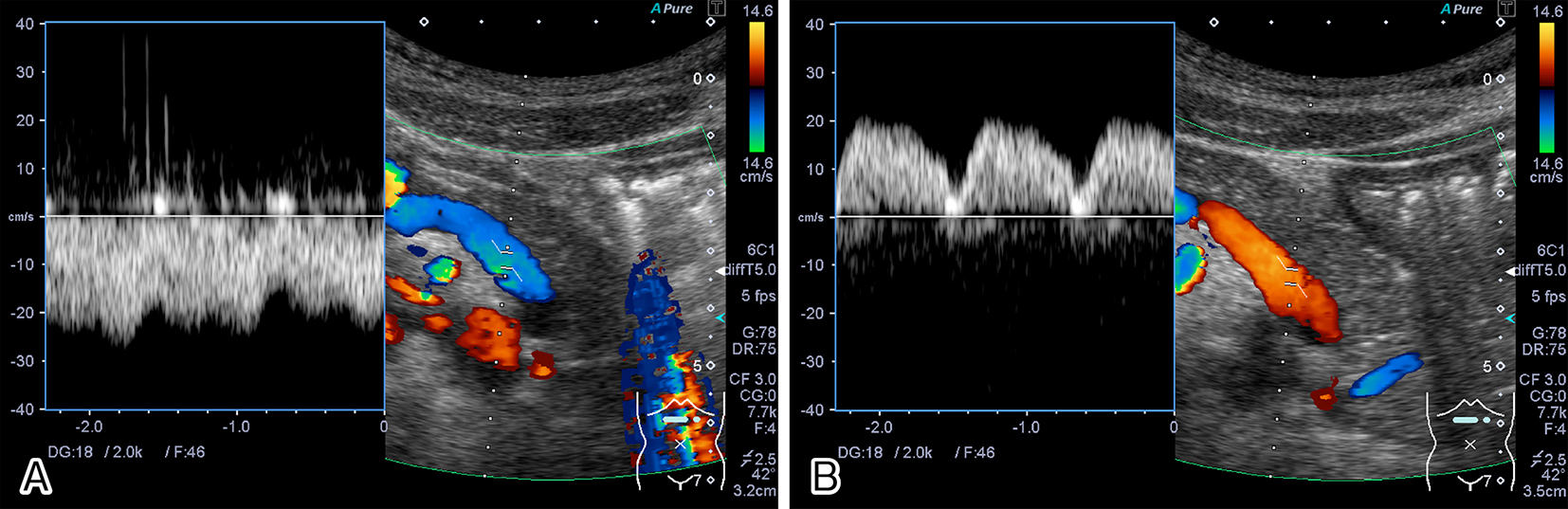
Figure 1. Color Doppler ultrasonographic images showing the splenic vein. (A) The splenic vein showed hepatofugal flow in the inspiratory phase. (B) The splenic vein showed hepatopetal flow in the expiratory phase.
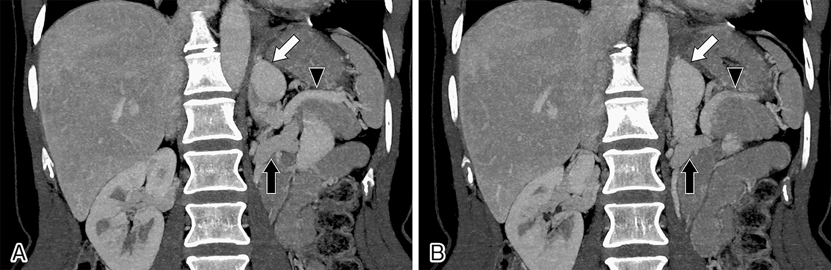
Figure 2. Computed tomographic scan images showing a splenorenal shunt (white arrows) between the splenic vein (black arrowheads) and the left renal vein (black arrows).