Corresponding author: Sho Kitagawa, bossa0405@yahoo.co.jp
DOI: 10.31662/jmaj.2018-0056
Received: December 6, 2018
Accepted: December 11, 2018
Advance Publication: February 1, 2019
Published: March 4, 2019
Cite this article as:
Kitagawa S, Minoura A. Congenital Splenorenal Shunt with Characteristic Ultrasonographic Findings. JMA J. 2019;2(1):91-92.
Key words: splenorenal shunt, congenital, ultrasonography, color Doppler imaging
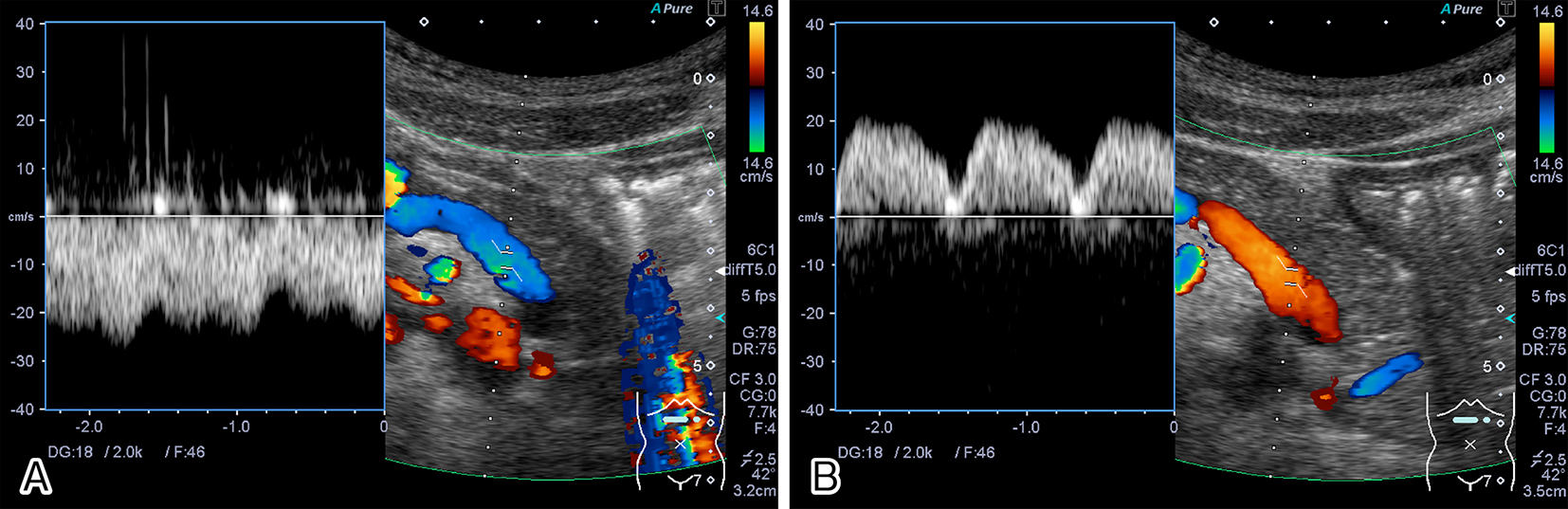
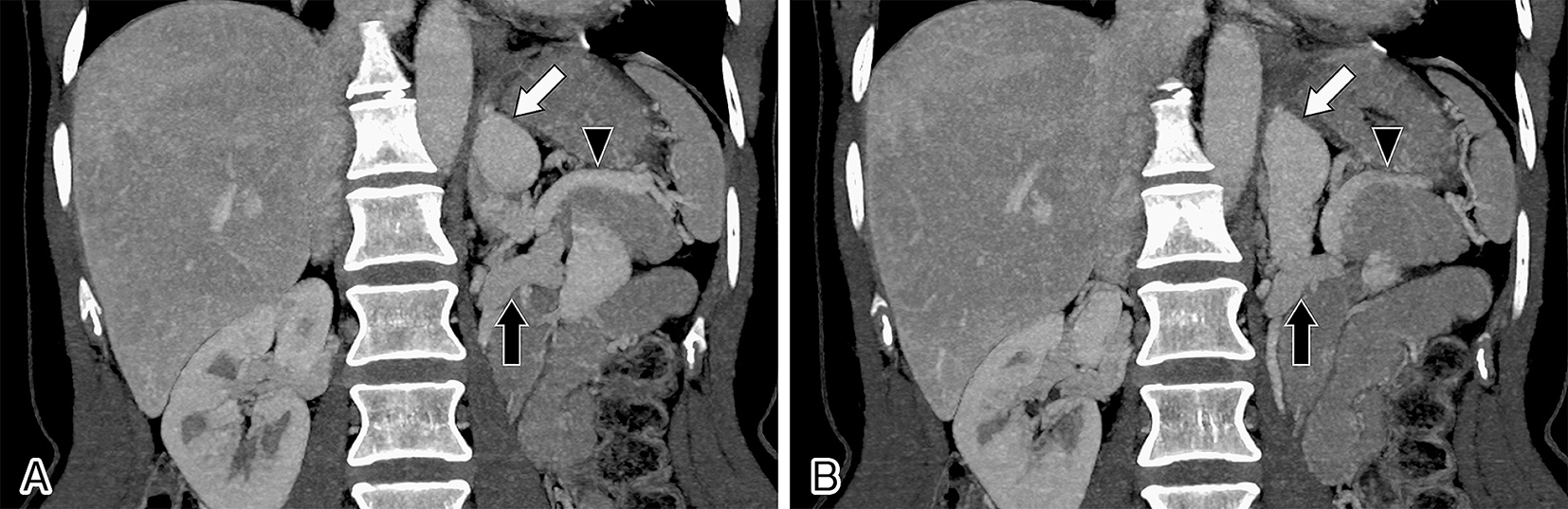
A 46-year-old woman presented for screening ultrasound examination. She had no history to suspect encephalopathy. During abdominal sonography, the direction of splenic venous flow was observed to be completely inverted in accordance with respiratory movements, namely, being hepatofugal in the inspiratory phase and hepatopetal in the expiratory phase (Figure 1). Subsequent computed tomography revealed a large tortuous vein between the splenic vein and the left renal vein (Figure 2). She had no history of trauma or surgery, and further examination showed no signs of portal hypertension. These findings were consistent with a diagnosis of congenital splenorenal shunt. Because her plasma ammonia levels were not elevated, the splenorenal shunt has been followed-up without any treatment. Congenital splenorenal shunt is a rare anomaly in patients without cirrhosis (1). In patients with portal hypertension, the direction of the splenic venous flow is to-and-fro or hepatofugal (2). In this case, the characteristic ultrasound findings were helpful for the diagnosis of splenorenal shunt.
None
SK wrote the manuscript. AM edited the manuscript.
We have obtained informed consent for this case report.
Lin YT, Chang CH, Chen WC. Asymptomatic congenital splenorenal shunt in a noncirrhotic patient with a left adrenal aldosterone-producing adenoma. Kaohsiung J Med Sci. 2009;25(12):669-74.
Yamada M, Ishida H, Komatsuda T, et al. Portal systemic shunt through the renal vein. Abdom Imaging. 2006;31(6):701-5.
