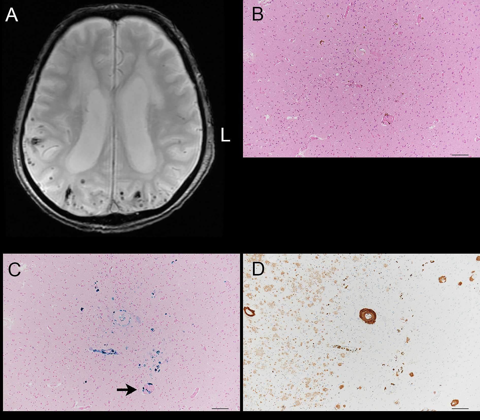
Figure 2. Images from a 90-year-old male with cerebral subcortical hemorrhage.
A. T2-weighted gradient echo images on cranial magnetic resonance imaging. Cerebral microbleeds (CMBs) can be seen in the temporal and occipital cortices.
B. Hematoxylin and eosin stain. Deposits of hemosiderin can be seen in the occipital lobe cortex. The accumulation of the hemosiderin deposits probably correspond to one CMB. Bar = 100 µm.
C. Berlin blue stain. Deposits of iron are present in the perivascular regions (arrow). Bar = 100 µm.
D. The vascular wall within the CMB was immunoreactive to monoclonal antibody raised against amyloid-β (11–28). Amyloid-β immunoreactive diffuse plaques are also present. Bar = 100 µm.
From: Cerebral Microbleeds Detected Using 3.0T Magnetic Resonance Imaging in 2,003 Patients with Ischemic or Hemorrhagic Stroke