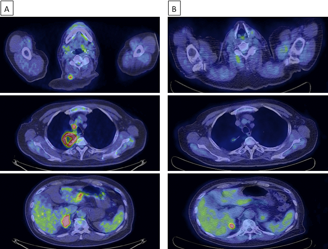
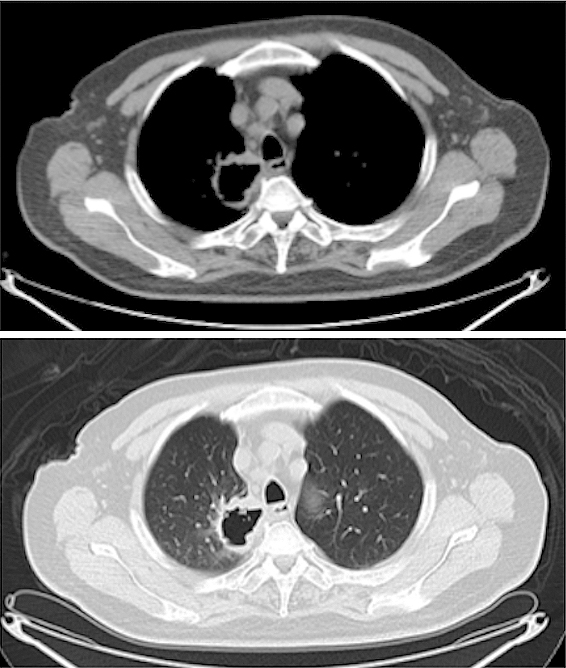
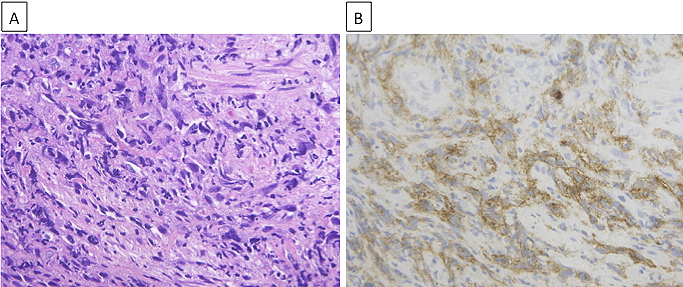
From: A Remarkable Clinical Response to Pembrolizumab in a Rare Spindle Cell Carcinoma of the Lung
From: A Remarkable Clinical Response to Pembrolizumab in a Rare Spindle Cell Carcinoma of the Lung
From: A Remarkable Clinical Response to Pembrolizumab in a Rare Spindle Cell Carcinoma of the Lung