Figure 1. Schematic representation of heart development and cardiac differentiation.
In the blastocyst stage, inner cell mass (ICM, blue) gives rise to three germ layers: ectoderm, endoderm, and mesoderm, and generates definitive structure of fetus. At embryonic day 6.5 (E6.5), endoderm and mesoderm layers emerge from primitive streak and migrate bilaterally. At E7.0, mesodermal cells migrate into the anterior region and generate cardiac crescent. At E7.5, first heart field (FHF, red) and second heart field (SHF, pink) are formed. By E9.5, looped heart tube is formed and outflow tract (OFT), right ventricle (RV) and left ventricle (LV), proepicardial organ (PEO, purple), etc. are formed. LV is derived from FHF, OFT and RV are mainly derived from SHF, and both the aria are derived from FHF and SHF. The colors are correlated with the stages in the cardiac differentiation.
From: Metabolic Regulation of Cardiac Differentiation and Maturation in Pluripotent Stem Cells: A Lesson from Heart Development
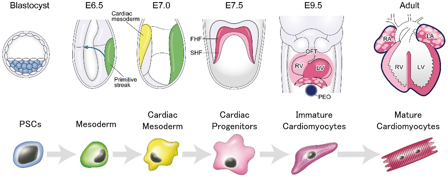
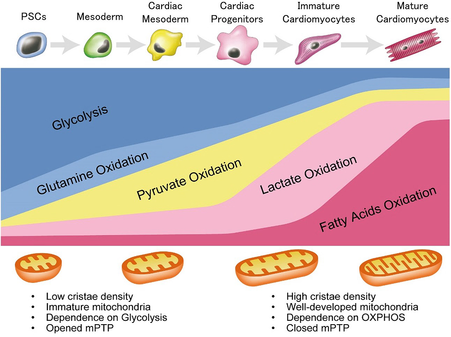
Figure 2. Stage-specific metabolic feature during cardiac differentiation from pluripotent stem cells.
PSCs mostly depend on glycolysis and glutamine oxidation metabolism. They exhibit immature mitochondria with low cristae density. During mesoderm and cardiac mesodermal stage, these cells still mainly depend on glycolysis and glutamine oxidation; however, pyruvate oxidation metabolism is also activated as mitochondrial development proceeds. In cardiac progenitor stage, these cells depend on aerobic and anaerobic metabolism equally. After differentiation into cardiomyocytes, mitochondrial number is increased and mitochondrial maturation is proceeded. Mitochondrial permeability transition pore (mPTP) is closed, when cardiac differentiation proceeded. mPTP closure enables the mitochondrial elongation and induces higher mitochondrial membrane potential. These cardiomyocytes depend mainly on lactate oxidation metabolism and fatty acid oxidation metabolism.
From: Metabolic Regulation of Cardiac Differentiation and Maturation in Pluripotent Stem Cells: A Lesson from Heart Development