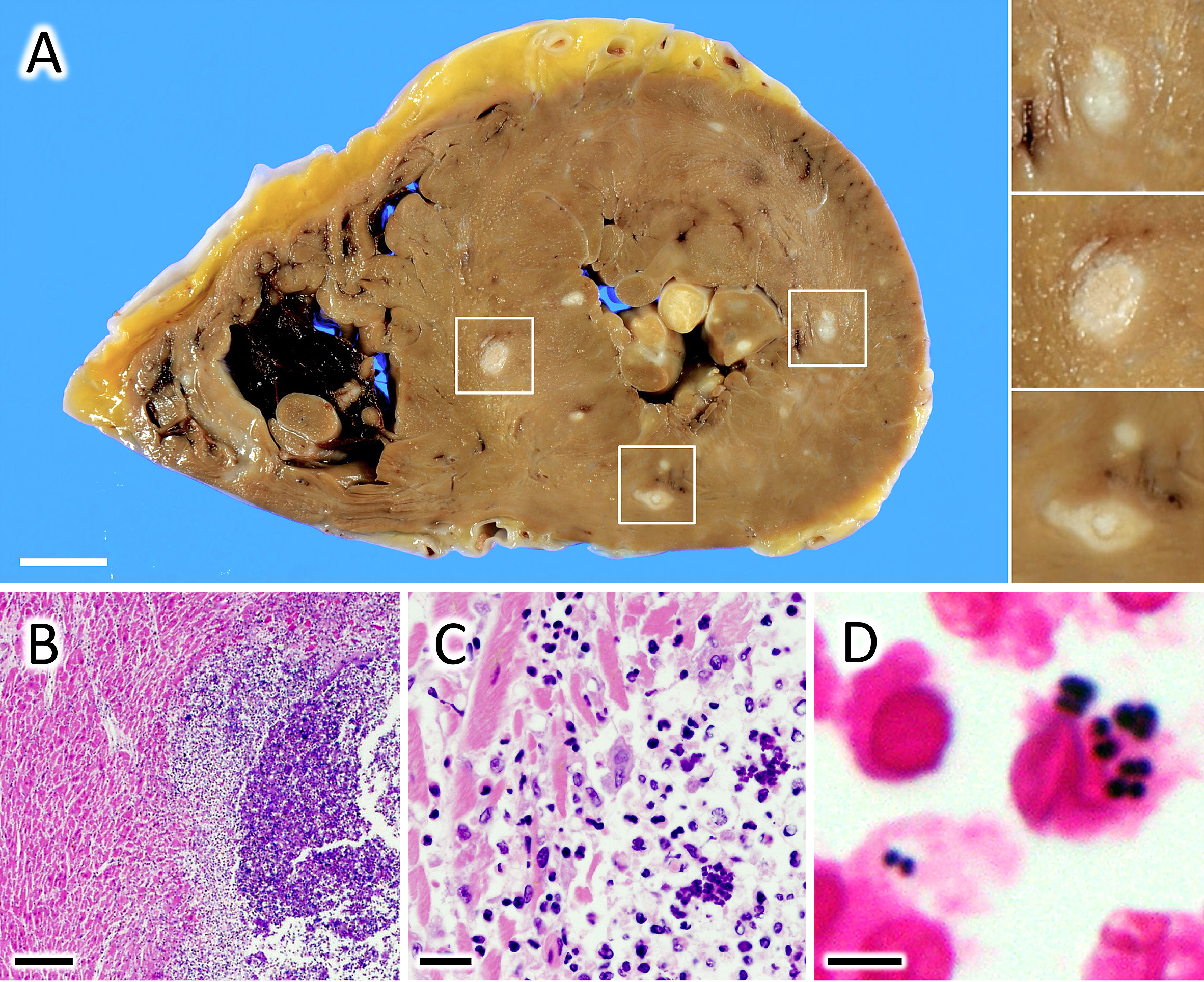
Figure 1. (A) Gross images of the heart. Multiple white nodules ranging in diameter from 1 to 10 mm are observed in the myocardium. Bar, 10 mm. (B-D) Microscopic images of hematoxylin and eosin (H & E; B, C) and Gram (D) staining of the white nodules. These are abscesses filled with numerous gram-positive diplococci and neutrophils. Bars, 500 μm (B), 50 μm (C), 5 μm (D).
From: Bacterial Myocarditis in a Patient with Cancer