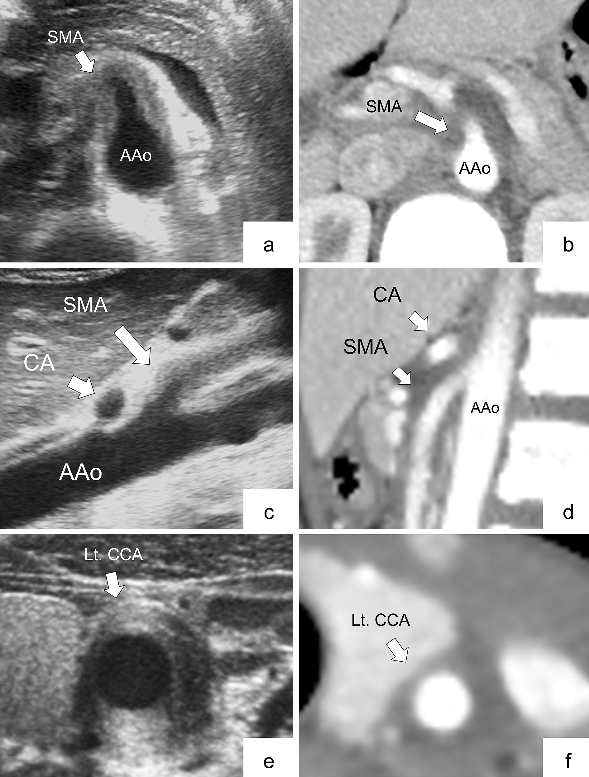
Figure 1. Representative images of arterial involvement comparing ultrasonography with contrast-enhanced computed tomography (CECT) in Patient No. 3 (panels a, b, c, d, e, and f).
a: Transverse view shows wall thickening at the level of the trunk of the superior mesenteric artery (SMA) in the ultrasonogram (arrow).
b: CECT at the same level of the abdominal ultrasonogram demonstrates wall thickening of the SMA (arrow).
c: Longitudinal view shows arterial stenosis at the level of the trunk of the celiac artery (CA), and wall thickening of the SMA in the ultrasonogram (arrow).
d: CECT at the same level of the abdominal ultrasonogram recognizes both findings of the CA and SMA (arrow).
e: Wall thickening of the left common carotid artery (CCA) is shown in the transverse view in the ultrasonogram (arrow).
f: Wall thickening of the left CCA is revealed by CECT at the same level of the neck ultrasonogram (arrow).
AAo, abdominal aorta; CA, celiac artery; CCA, common carotid artery; Lt., left; SMA, superior mesenteric artery.
From: Ultrasonography as a Diagnostic Support Tool for Childhood Takayasu Arteritis Referred to as Fever of Unknown Origin: Case Series and Literature Review