Figure 1. a. Abdominal precontrast computed tomography showing splenomegaly. b-d. Abdominal contrast-enhanced computed tomography showing multiple splenic hypodense lesions and no enlarged lymph nodes.

From: Primary Splenic Lymphoma: An Uncommon Cause of Persistent Fever Diagnosed by Examining the “Forgotten Organ”

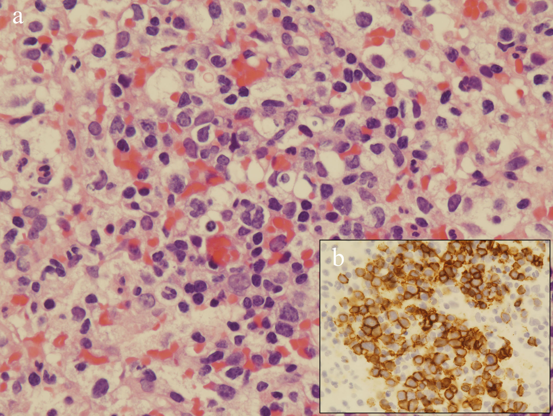
Figure 2. a. Microscopic examination of the splenic biopsy specimen showing diffuse proliferation of atypical cells (hematoxylin-eosin stain; original magnification, 400×). b. Immunohistochemical analysis showing positive CD20 (original magnification, 400×).

From: Primary Splenic Lymphoma: An Uncommon Cause of Persistent Fever Diagnosed by Examining the “Forgotten Organ”

PAGE TOP