Figure 1. (A) Computed tomography (CT) of the chest revealed multiple small nodular infiltrates with cavities (arrow).
(B) Abdominal CT revealed the enlargement of both the kidneys.

From: Rupture of Renal Artery Aneurysm in a Patient with Granulomatosis with Polyangiitis
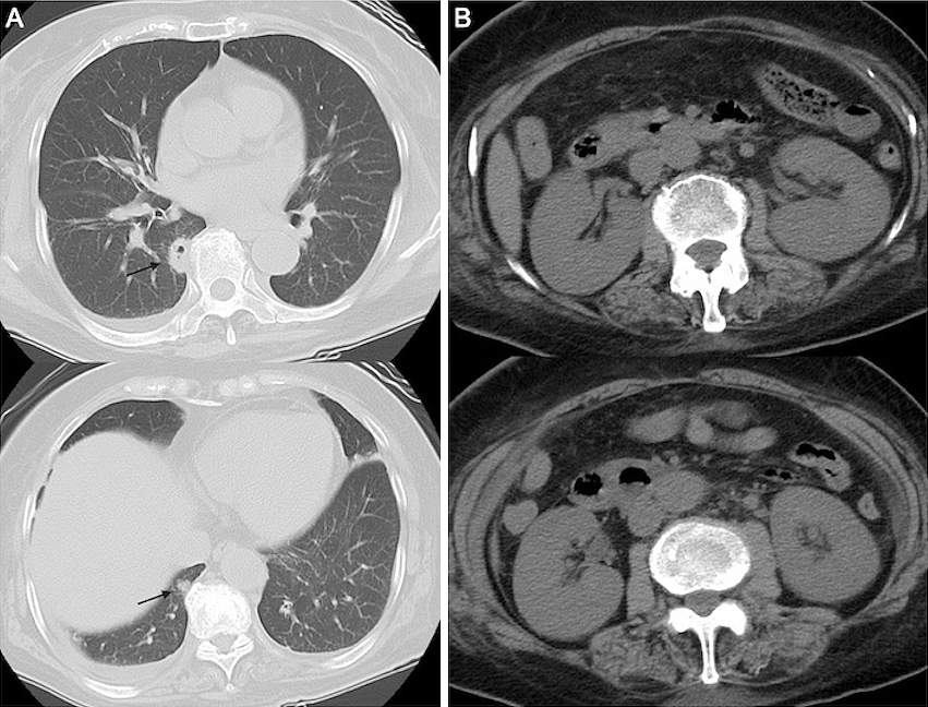
Figure 2. Images of computed tomography with abdominal hematoma. Contrast medium extravasation from the kidney (arrowhead) and aneurysm formation (arrow).

From: Rupture of Renal Artery Aneurysm in a Patient with Granulomatosis with Polyangiitis
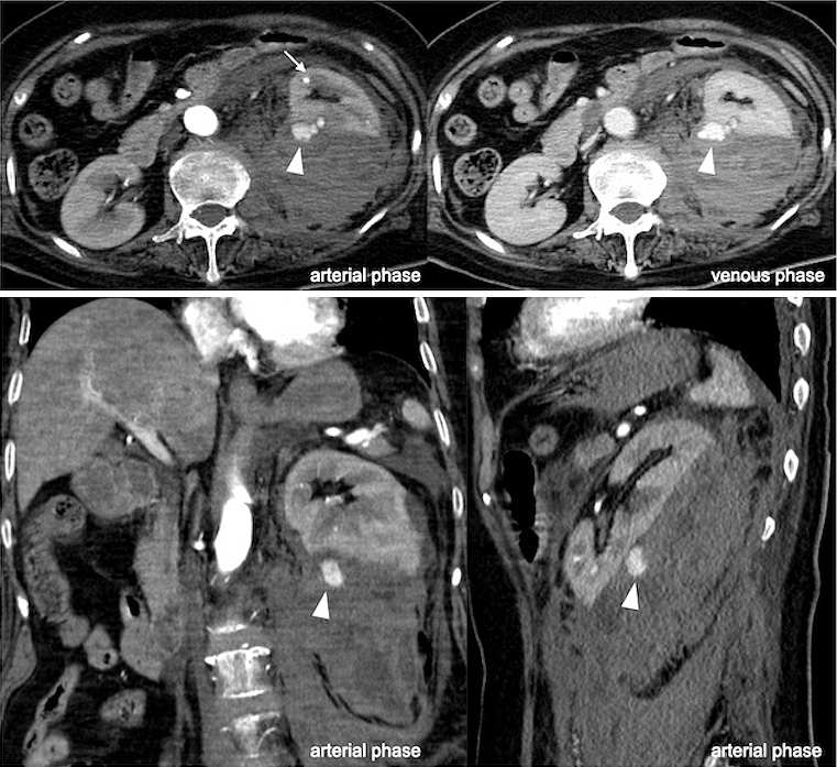
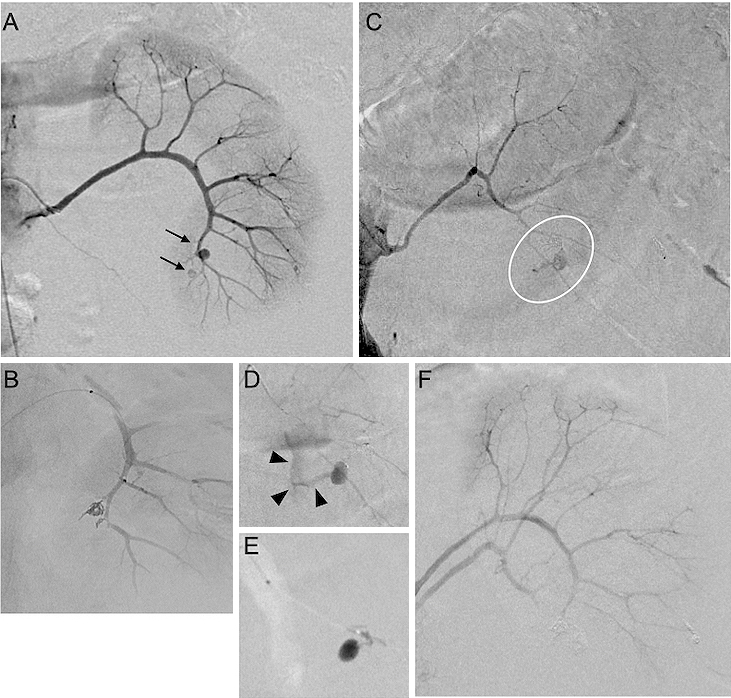
Figure 3. (A) Angiography revealed the aneurysms (arrow) from the anteroinferior segmental artery of the kidney.
(B) Coil embolization.
(C and D) Angiography showing the extravasation of contrast medium from the posterior segmental artery of the kidney.
(E) Injection of N-butyl cyanoacrylate.
(F) Completion of hemostasis.

From: Rupture of Renal Artery Aneurysm in a Patient with Granulomatosis with Polyangiitis
PAGE TOP