Figure 2. Light microscopy of the kidney showing diffuse mesangial hypercellularity (A, periodic acid-Schiff staining, 400×); spike formation (black arrows) and double contouring (white arrowheads) of the glomerular basement membrane (B, periodic acid-methenamine-silver staining, 800×); no arterial lesions (C, Masson’s trichrome staining, 400×); mild tubular atrophy (D, periodic acid-Schiff staining, 400×).
From: Chilblain Lupus Erythematosus
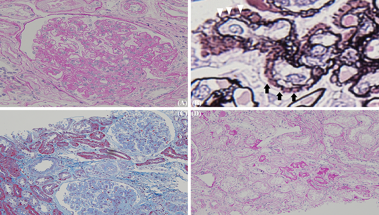
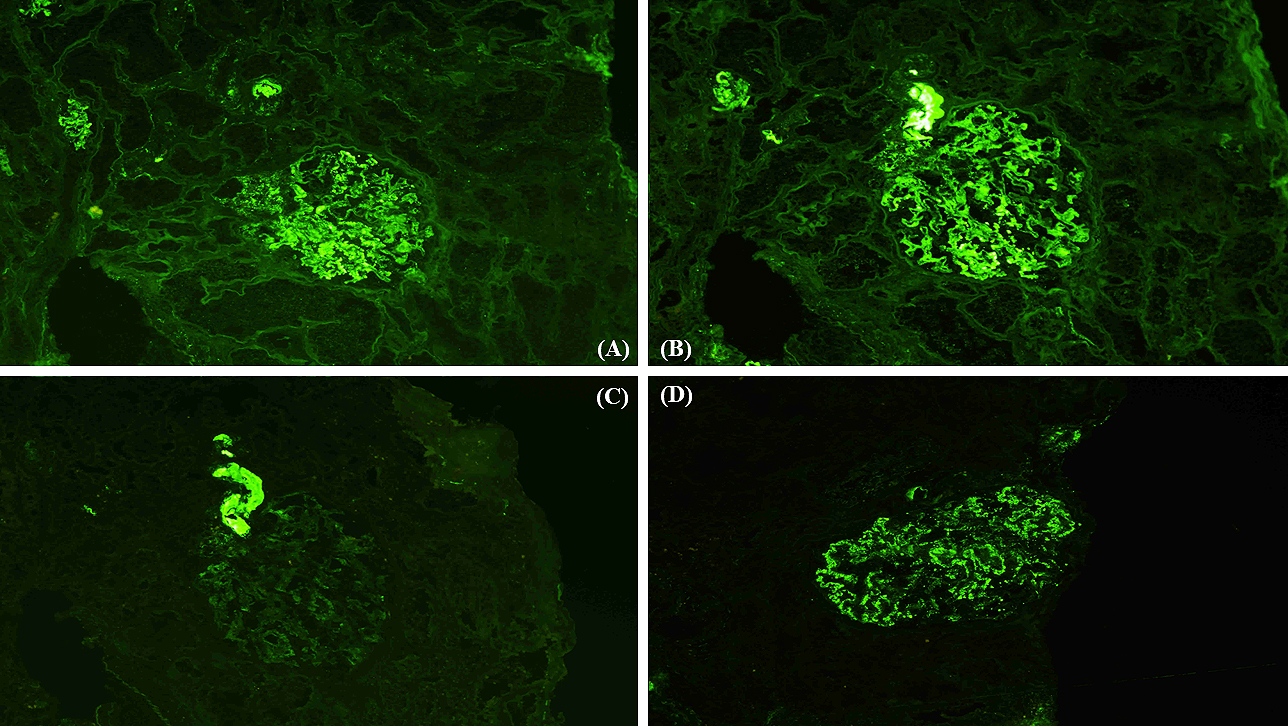
Figure 3. Immunofluorescence microscopy of the kidney showing the depositions of immunoglobulins G, A, and M, and C1q. (A) immunoglobulin G; (B) immunoglobulin A; (C) immunoglobulin M; (D) C1q, 400×.
From: Chilblain Lupus Erythematosus