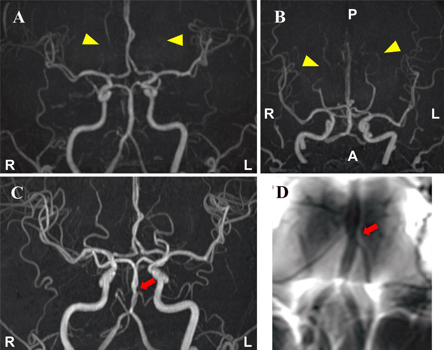
Figure 1. Magnetic resonance angiography (MRA) on day 3 after the onset revealed somewhat poor delineation of the bilateral distal posterior cerebral arteries (arrowheads), which suggested possible vasoconstriction (A, B). MRA on day 17 showed segmental constriction of the basilar artery (arrow) (C). Basi-parallel anatomical scanning on the same day revealed segmental constriction of the external vessel wall of the basilar artery (arrow) (D).
From: Migration of Vasoconstriction in Reversible Cerebral Vasoconstriction Syndrome