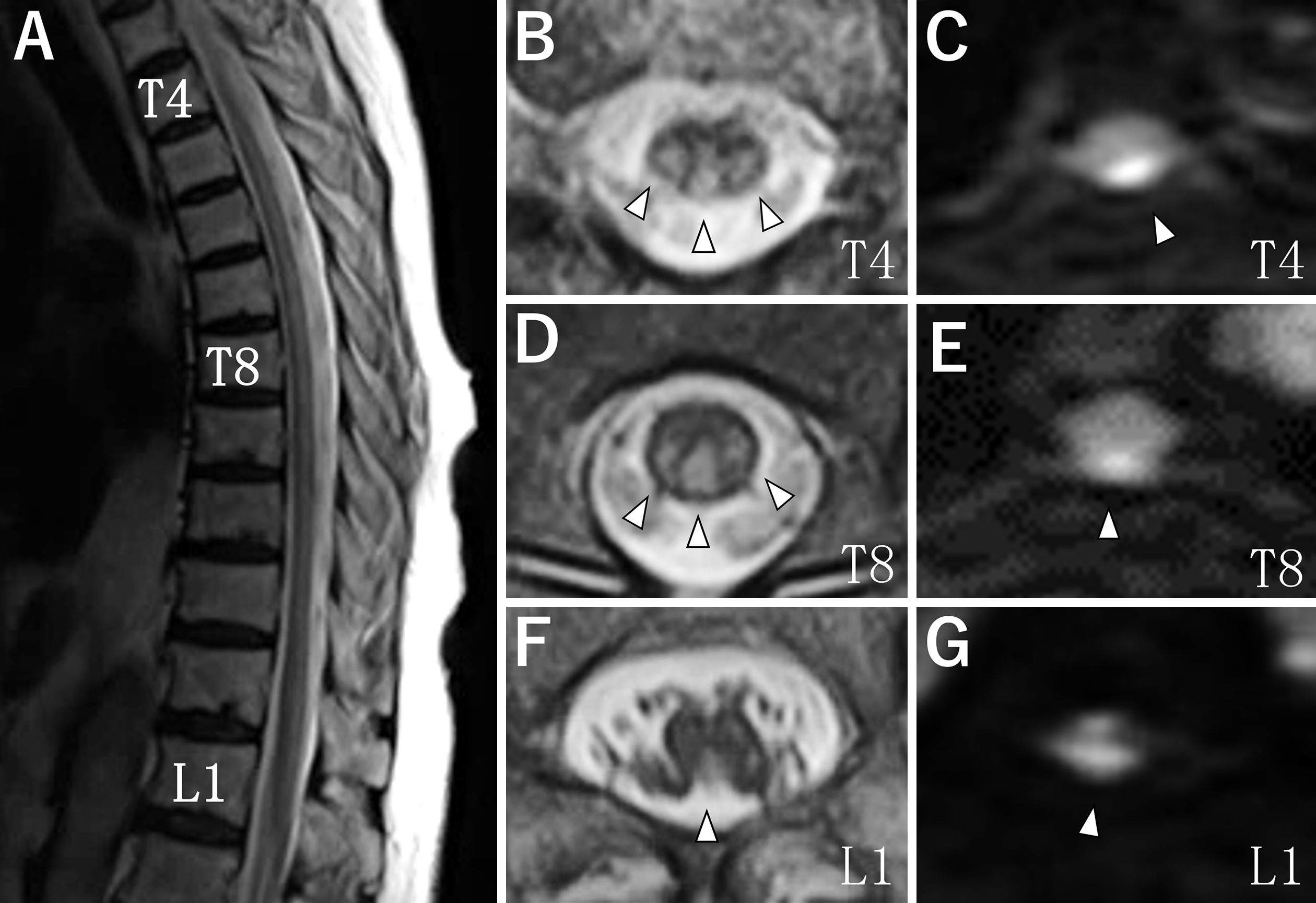
Figure 1. Spinal magnetic resonance imaging.
Sagittal T2-weighted imaging (T2WI) showed longitudinal hyperintensity from vertebral level T4 to the conus medullaris of the spinal cord (A). Axial images showed hyperintensity in the posterior part of the spinal cord on T2WI and diffusion-weighted imaging (DWI) (B-G arrowheads). T2WI (B, D, F). DWI (C, E, G). The vertebral levels of each image are as follows: T4 (B, C), T8 (D, E), and L1 (F, G).
From: Extremely Long Spinal Cord Infarction