Corresponding author: Tatsuya Tanaka, s96047@hotmail.com
DOI: 10.31662/jmaj.2023-0049
Received: March 23, 2023
Accepted: April 24, 2023
Advance Publication: June 12, 2023
Published: July 14, 2023
Cite this article as:
Tanaka T, Sashida R, Hirokawa Y, Matsuno A. Aberrant Right Subclavian Artery Identified before Carotid Artery Stenting. JMA J. 2023;6(3):352-353.
Key words: aberrant right subclavian artery, aortic arch anomaly, carotid artery stenting, retroesophageal right subclavian artery, three-dimensional computed tomography angiography
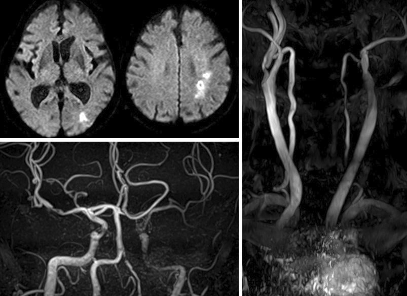
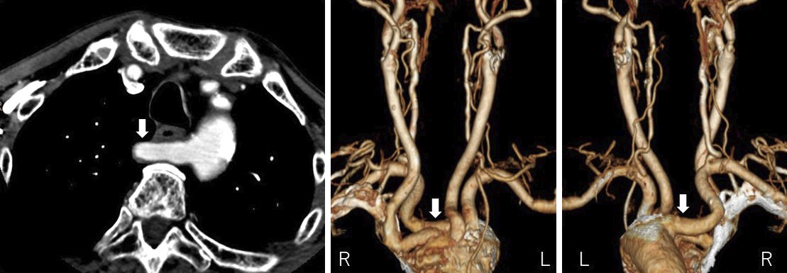
An 84-year-old man with right common iliac artery dissection presented with a complaint of transient right upper limb weakness. Magnetic resonance imaging of the head revealed a cerebral infarct in the left hemisphere, and magnetic resonance angiography revealed severe stenosis of the left internal carotid artery (Figure 1). Antithrombotic therapy was performed, and a transradial approach was selected as the patient had previously undergone iliac artery dissection. Preoperative three-dimensional computed tomography angiography revealed an aberrant right subclavian artery with a retroesophageal course originating from the most distal branch of the aortic arch (Figure 2, arrow). Carotid artery stenting was performed via the left femoral artery.
Aberrant right subclavian artery is a rare aortic arch anomaly, affecting only 0.4%-2.0% of the population (1). Most patients remain asymptomatic; however, aneurysmal dilatation, aortic dissection, and tracheal and esophageal compression are some serious vascular complications.
Recently, endovascular treatment via the radial or brachial artery has gained more popularity. Preoperative three-dimensional computed tomography angiography alerts physicians about any morphological abnormalities and reduces surgical risks.
None
TT wrote the first draft and managed the submission process. RS, YH, and AM commented on previous versions of the manuscript. All authors read and approved the final version of the manuscript.
We have obtained informed consent for this case report.
Freed K, Low VH. The aberrant subclavian artery. AJR Am J Roentgenol. 1997;168(2):481-4.
