Corresponding author: Naoya Fujita, raoh0615@gmail.com
DOI: 10.31662/jmaj.2022-0223
Received: December 20, 2022
Accepted: January 26, 2023
Advance Publication: March 24, 2023
Published: April 14, 2023
Cite this article as:
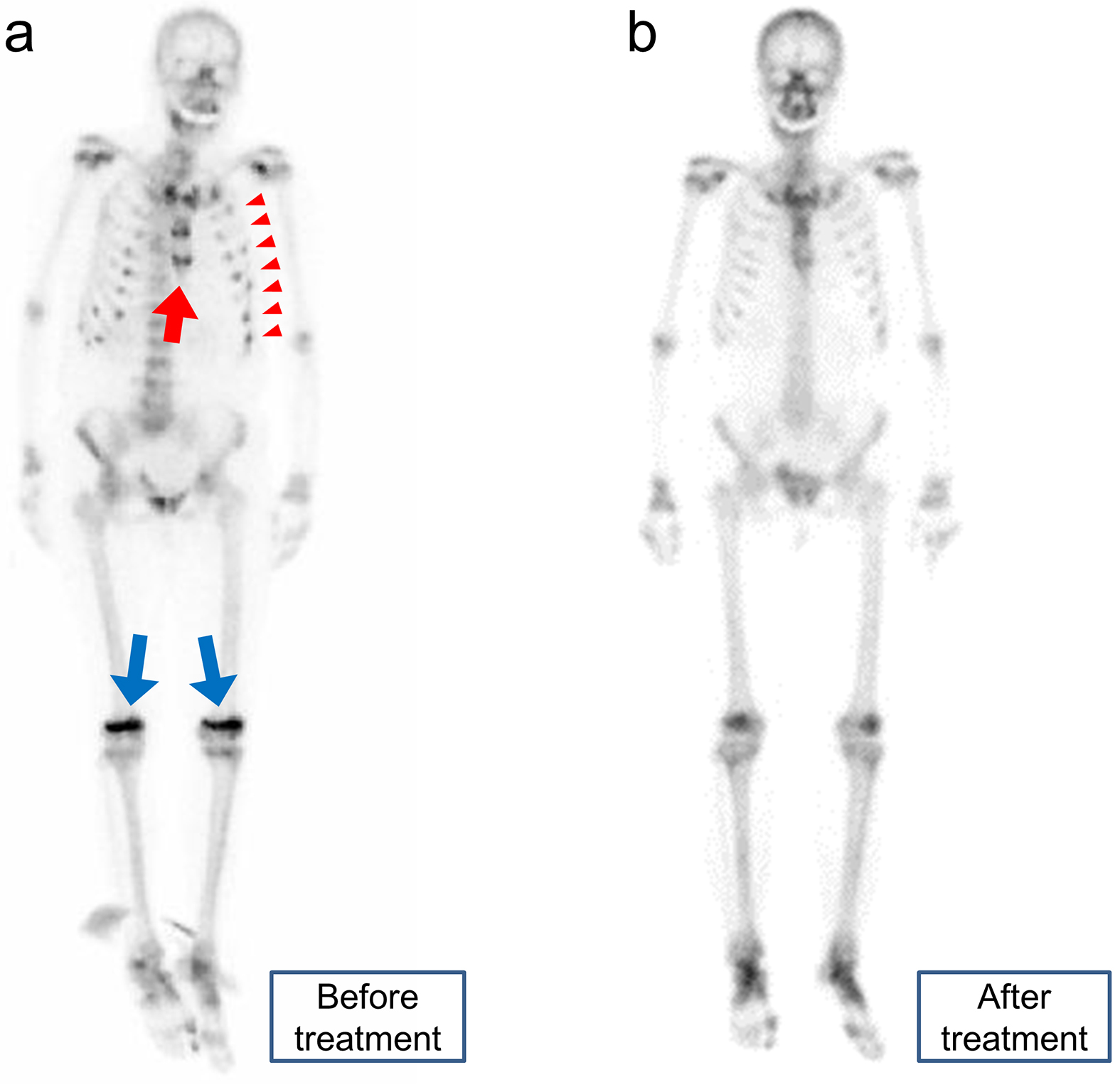
Fujita N, Ono Y. Scintigraphic Findings in FGF23-related Hypophosphatemic Osteomalacia. JMA J. 2023;6(2):214-215.
Key words: FGF23-related hypophosphatemic osteomalacia, bone scintigraphy, tie sign, rachitic rosary sign
A 67-year-old woman presented with systemic bone pain and muscle weakness. Laboratory examinations revealed high levels of serum alkaline phosphatase, normal corrected calcium, and low inorganic phosphate levels. Bone scintigraphy revealed multiple uptake in the sternum, costochondral junction, and knee joints, which were known as the “tie sign,” “rachitic rosary sign,” and “accumulation in the knees,” respectively (Figure 1a) (1), (2), (3). These signs are the characteristic of fibroblast growth factor 23 (FGF23)-related hypophosphatemic osteomalacia, and the serum FGF23 levels were high (184 pg/mL; normal range: 19.9-52.9 pg/mL). Although she was suspected to have tumor-induced osteomalacia, systemic venous samplings of FGF23 and 111In-octreotide somatostatin receptor scintigraphy were unable to identify the tumor. Her symptoms and multiple uptake on bone scintigraphy almost disappeared after initiating active vitamin D and phosphate replacement (Figure 1b). Although bone scintigraphy is not useful for localizing tumors, it is recommended to differentiate the etiology of bone pain.
None
Naoya Fujita wrote the first draft. Yosuke Ono suggested improvements and revised the manuscript.
Written informed consent was obtained from the patient to publish this report in accordance with the journal’s patient consent policy.
Chakraborty PP, Bhattacharjee R, Mukhopadhyay S, et al. 'Rachitic rosary sign' and 'tie sign' of the sternum in tumour-induced osteomalacia. BMJ Case Rep. 2016;bcr2016214766.
Liu SZ, Zhou X, Song A, et al. Rachitic rosary sign and tie sign in tumour-induced osteomalacia. QJM. 2020;113(4):284-5.
Yamamoto K, Honda H, Ota I, et al. Triad signs shown by bone scintigraphy in FGF23-related osteomalacia. QJM. 2022;114(12):887-8.
