Corresponding author: Hiroshi Shiba, hshiba0124@gmail.com
DOI: 10.31662/jmaj.2023-0028
Received: February 24, 2023
Accepted: March 27, 2023
Advance Publication: May 22, 2023
Published: July 14, 2023
Cite this article as:
Shiba H, Endo T, Fujikawa H, Tsukamoto T. Giant Rectus Sheath Hematoma: Pseudobladder Sign. JMA J. 2023;6(3):348-349.
Key words: rectus sheath hematoma, COVID-19, anticoagulation therapy, ultrasound
A 75-year-old woman with coronavirus disease 2019 (COVID-19) and on prophylactic heparin reported acute abdominal pain provoked by coughing on Day 6 of hospitalization. She developed peritoneal signs and hemorrhagic shock on Day 7. A bedside ultrasound revealed a hypoechoic fluid collection over a hyperechoic layer, which was initially thought to be a urinary bladder hematoma (Figure 1A). However, dynamic contrast-enhanced computed tomography revealed rectus sheath hematoma (RSH) with multiple areas of active extravasation (Figure 1B and 1C). The patient was conservatively treated and underwent elective percutaneous drainage with no recurrence. Interventional radiology embolization was avoided due to the risk of tissue necrosis.
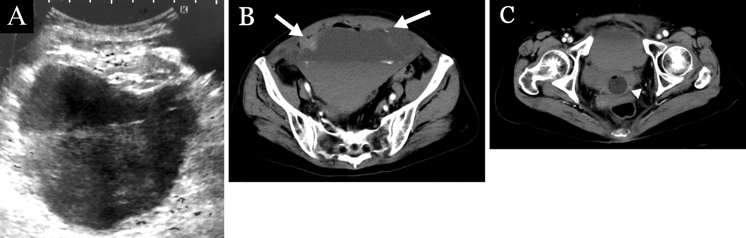
RSH should be suspected in COVID-19 patients with abdominal pain, as anticoagulation, cough, and COVID-19 infection itself can be risk factors for RSH (1), (2). Although ultrasound is commonly used as the first-line diagnostic modality for RSH, it may lead to misdiagnosis when it fails to detect the origin of a large hematoma (3). When the hematoma extends to the pelvic area, it may mimic bladder distension, as observed in our case. To alert clinicians of this misleading image, we propose to refer it as the “pseudobladder sign.”
None
HS acquired data and drafted the manuscript. TE, HF, and TT reviewed and supervised the manuscript.
We have obtained informed consent for this manuscript.
In this study, IRB approval was not required.
Cherry WB, Mueller PS. Rectus sheath hematoma: review of 126 cases at a single institution. Medicine. 2006;85(2):105-10.
Özer M, Terzioglu SG, Keskinkılıç Yağız B, et al. Does COVID-19 increase the incidence of spontaneous rectus sheath hematoma? Ulus Travma Acil Cerrahi Derg. 2022;28(6):920-6.
Bello G, Blanco P. Giant rectus sheath hematoma. Ultrasound J. 2019;11(1):13.
